Copyright
©The Author(s) 2015.
World J Gastroenterol. Apr 7, 2015; 21(13): 4020-4029
Published online Apr 7, 2015. doi: 10.3748/wjg.v21.i13.4020
Published online Apr 7, 2015. doi: 10.3748/wjg.v21.i13.4020
Figure 1 Preoperative magnetic resonance imaging of the perianal region and its schematic diagram showing a recurrent horseshoe abscess and fistula from 2 to 10 o’clock position in a 29-year-old female patient.
There was no external opening and the internal opening was at the posterior midline. Arrows in the upper pictures show the position of the horseshoe abscess.
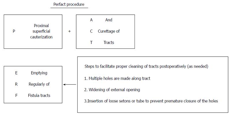
Figure 2 PERFACT procedure - an overview.
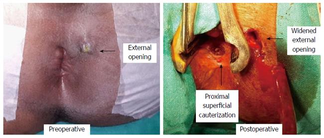
Figure 3 Management of a 45-year-old male patient by PERFACT procedure.
He had a recurrent transsphincteric fistula with external opening at 2 o’clock and internal opening at 6 o’clock posterior midline. Shows proximal superficial cauterization and widening of the external opening. Horizontal arrows show the external opening - preoperative in the left picture and postoperative in the right. The vertical arrow in the right picture shows the position of proximal superficial cauterization.
Figure 4 Management of a 29-year-old female patient by PERFACT procedure.
She had a horseshoe abscess and fistula from 2 to 10 o’clock. There was no external opening and the internal opening was at the posterior midline. The right bottom picture shows complete healing of the fistula (MRI and diagram of this patient shown in Figure 2).
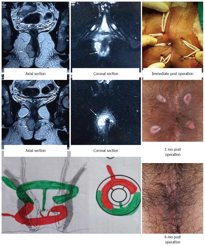
Figure 5 Management of a 22-year-old male patient by PERFACT procedure.
He had a double horseshoe intersphincteric abscess and fistula. External opening is at 11 o’clock and the internal opening was not traceable intraoperatively. Electrocauterization was done at both anterior and posterior midline. The left upper picture shows the MRI and the right bottom picture was taken after the final cure (posterior horseshoe abscess shown by green color was at a higher level). Arrows in the upper left pictures show the position of the horseshoe abscess and tracts.
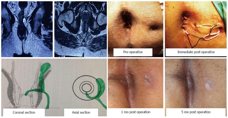
Figure 6 Management of a 36-year-old male patient by PERFACT procedure.
He had a supralevator extension at 3 o’clock. External opening is at 3 o’clock and internal opening at 6 o’clock posterior midline. The left upper picture shows the MRI (arrows show the supralevator extension) and the right bottom picture was taken after the patient was fully cured.
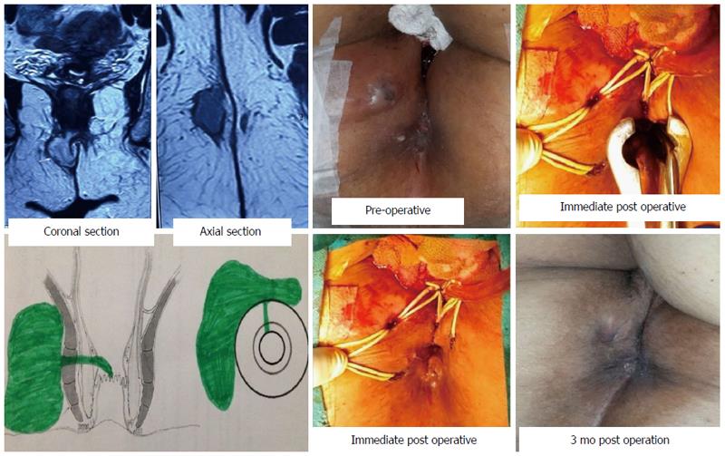
Figure 7 Management of a 55-year-old female patient by PERFACT procedure.
She had a large anterior abscess and a fistula. External opening is at 11 o’clock and internal opening at 12 o’clock anterior midline. The left upper picture shows the MRI and the right bottom picture is taken after the final cure.
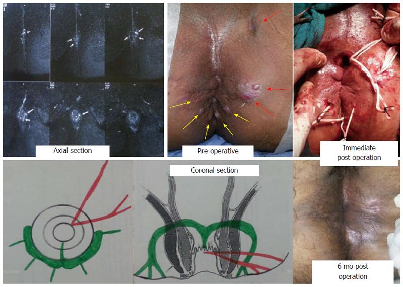
Figure 8 Management of a 38-year-old male patient by PERFACT procedure.
He had a total of eight external openings and tracts (including a horseshoe one). The left upper picture shows the MRI and the right bottom picture is taken after the final cure. Arrows in the upper left picture show the position of the multiple tracts and the upper middle picture shows the multiple external openings.
- Citation: Garg P, Garg M. PERFACT procedure: A new concept to treat highly complex anal fistula. World J Gastroenterol 2015; 21(13): 4020-4029
- URL: https://www.wjgnet.com/1007-9327/full/v21/i13/4020.htm
- DOI: https://dx.doi.org/10.3748/wjg.v21.i13.4020


