Copyright
©The Author(s) 2015.
World J Gastroenterol. Mar 14, 2015; 21(10): 2973-2981
Published online Mar 14, 2015. doi: 10.3748/wjg.v21.i10.2973
Published online Mar 14, 2015. doi: 10.3748/wjg.v21.i10.2973
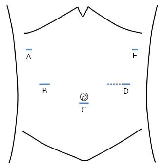
Figure 1 Placement of the surgical ports and mini-laparotomy.
A: 5 mm; B: 5 mm; C: 12 mm (the working port); D: 10 mm (the camera port); E: 5 mm (the site of the mini-laparotomy, dotted line).
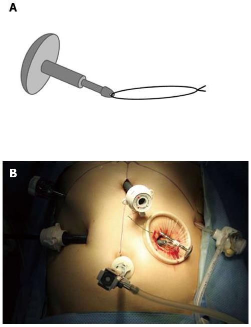
Figure 2 Anvil preparation.
A: The stay-suture is applied to the tip of the anvil; B: The anvil is inserted into the abdominal cavity through the mini-laparotomy.
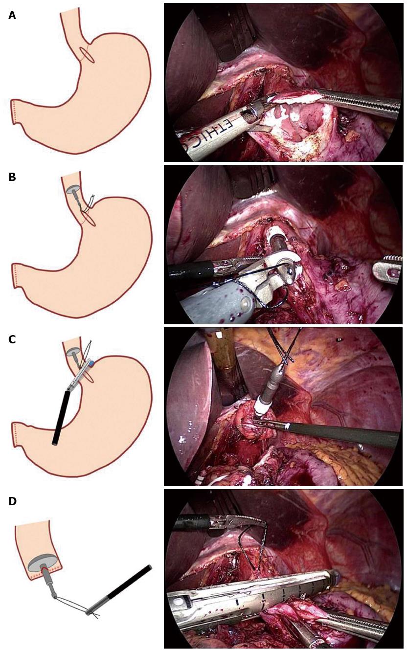
Figure 3 Anvil insertion.
A: A small gastrotomy (approximately 3 cm) is performed across the esophagogastric junction using a harmonic scalpel; B: the prepared anvil is inserted into the esophagus through the gastrotomy until it reaches the thoracic esophagus; C: The distal esophagus is transected with a linear stapling. To avoid disrupting the stay-suture, the stay-suture is lifted cranially; D: The anvil is completely placed in the esophageal stump after the stay-suture is pulled out.
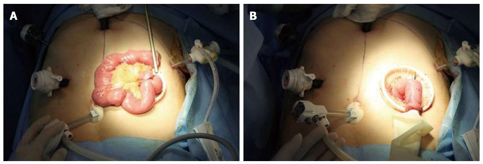
Figure 4 Jejunojejunostomy.
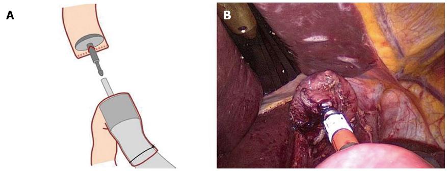
A: An extracorporeal end-to-side jejunojejunostomy is manually performed 45 cm below the site at which the esophagojejunostomy will be performed; B: For intracorporeal use, a surgical glove is attached to the circular stapler. The circular stapler is inserted into the Roux limb, and the Roux limb is tied to the body of the circular stapler to prevent separation from the circular stapler.
Figure 5 Intracorporeal end-to-side esophagojejunostomy.
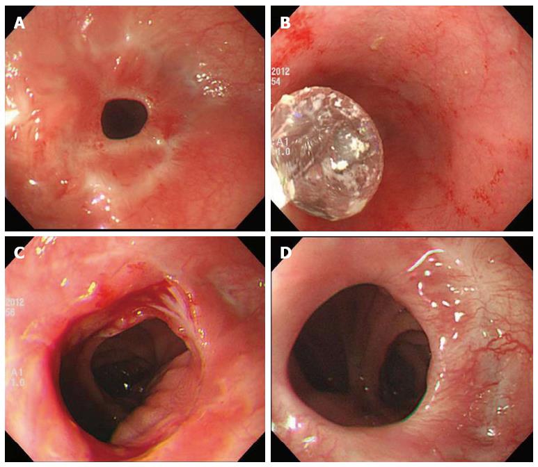
Figure 6 Anastomotic stricture present after discharge.
A: Before intervention; B: Interventional endoscopic ballooning; C: Immediate state after intervention; D: Appearance after 4 mo.
- Citation: Kim JH, Choi CI, Kim DI, Kim DH, Jeon TY, Kim DH, Park DY. Intracorporeal esophagojejunostomy using the double stapling technique after laparoscopic total gastrectomy: A retrospective case-series study. World J Gastroenterol 2015; 21(10): 2973-2981
- URL: https://www.wjgnet.com/1007-9327/full/v21/i10/2973.htm
- DOI: https://dx.doi.org/10.3748/wjg.v21.i10.2973


