Copyright
©The Author(s) 2015.
World J Gastroenterol. Jan 7, 2015; 21(1): 369-372
Published online Jan 7, 2015. doi: 10.3748/wjg.v21.i1.369
Published online Jan 7, 2015. doi: 10.3748/wjg.v21.i1.369
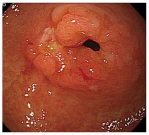
Figure 1 Upper gastrointestinal endoscopic examination.
Superficial slightly depressed-type tumor [cT1b (UICC)] at the pyloric region of gastric tube was diagnosed as adenocarcinoma by endoscopic biopsy.
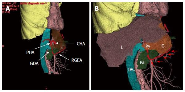
Figure 2 Three-dimensional computed tomography.
A: The courses of the branches of celiac artery were well visualized while obtaining surgical navigation; B: Relationship with the gastric tube and surrounding structures were well visualized while obtaining surgical navigation. Arrows shows the running of right gastroepiploic artery. L: Liver, IVC: Inferior vena cava; Pa: pancreas; G: Gastric tube; Py: Pylorus; CHA: Common hepatic artery; RGEA: Right gastroepiploic artery; GDA: Gastroduodenal artery; PHA: Proper hepatic artery.
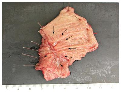
Figure 3 Resected specimen of the gastric tube.
Resected specimen showed a slightly depressed-type tumor (arrows) in the pyloric lesion of gastric tube measuring 20 mm × 15 mm in size.
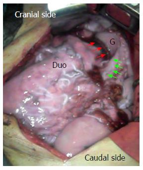
Figure 4 Intraoperative indocyanine green fluorescence imaging.
Intravenous administration of indocyanine green (ICG) showed that the blood flow of the right gastroepiploic artery and surgical margin of the gastric tube was sufficiently maintained in the proximal gastric tube. Proximal margin of gastric tube (red arrows); Course of the right gastroepiploic artery (green arrows). Duo: Margin of the duodenum; G: Proximal gastric tube.
- Citation: Nakano T, Sakurai T, Maruyama S, Ozawa Y, Kamei T, Miyata G, Ohuchi N. Indocyanine green fluorescence and three-dimensional imaging of right gastroepiploic artery in gastric tube cancer. World J Gastroenterol 2015; 21(1): 369-372
- URL: https://www.wjgnet.com/1007-9327/full/v21/i1/369.htm
- DOI: https://dx.doi.org/10.3748/wjg.v21.i1.369


