Copyright
©The Author(s) 2015.
World J Gastroenterol. Jan 7, 2015; 21(1): 360-368
Published online Jan 7, 2015. doi: 10.3748/wjg.v21.i1.360
Published online Jan 7, 2015. doi: 10.3748/wjg.v21.i1.360
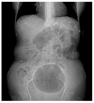
Figure 1 Abdominal X-ray shows a large gas-filled cavity in the lower abdomen.
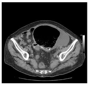
Figure 2 Axial non-contrast computed tomography scan shows a 15.
5 cm x 10.5 cm cystic lesion containing air and fluid, with a thick wall.
- Citation: Nigri G, Petrucciani N, Giannini G, Aurello P, Magistri P, Gasparrini M, Ramacciato G. Giant colonic diverticulum, clinical presentation, diagnosis and treatment: Systematic review of 166 cases. World J Gastroenterol 2015; 21(1): 360-368
- URL: https://www.wjgnet.com/1007-9327/full/v21/i1/360.htm
- DOI: https://dx.doi.org/10.3748/wjg.v21.i1.360


