Copyright
©2014 Baishideng Publishing Group Co.
World J Gastroenterol. Feb 28, 2014; 20(8): 2030-2041
Published online Feb 28, 2014. doi: 10.3748/wjg.v20.i8.2030
Published online Feb 28, 2014. doi: 10.3748/wjg.v20.i8.2030
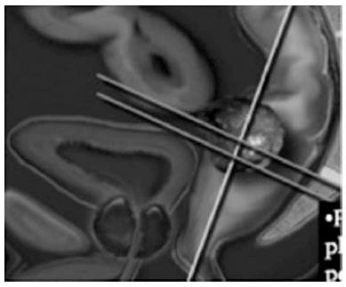
Figure 1 Magnetic resonance imaging technique.
T2 weighted non-fat suppressed sequence in all three orthogonal planes to tumor axial, coronal, sagittal should be used.
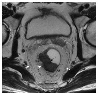
Figure 2 T3 tumor, circumferential resection margin not threatened.
T2W axial magnetic resonance imaging image shows a mildly hyperintense proliferative tumor along the right lateral and posterior wall (black arrow). Arrow head shows the tumor reaching upto the muscularis with spiculation in the adjacent perirectal fat. The white arrows show the mesorectal fascia which is not involved/threatened.
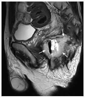
Figure 3 Mucinous adenocarcinoma of rectum.
Sagittal T2 weighted MRI image showing a circumferential rectal tumour with high signal intensity (arrows) characteristic of a mucinous tumor. MRI: Magnetic resonance imaging.
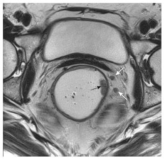
Figure 4 T1 N+ tumor.
T2W axial magnetic resonance image shows two small nodes in the mesorectal fat on the left (white arrows) with irregular borders and signal intensity similar to primary tumor along left lateral wall (black arrow).
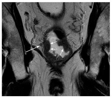
Figure 5 T2 tumor coronol views with extramural venous invasion.
Coronal T2W magnetic resonance imaging shows a cicumferential tumor in the rectum (small arrows) reaching the muscularis with extramural venous invasion (long arrow).
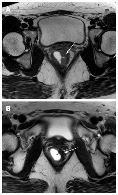
Figure 6 (A) Pre-chemoradiotherapy and (B) postchemoradiotherapy status.
Arrows point to tumor along left lateral wall tumor which is mildly hyperintense in A that has become darkly hypointense in B indicating response (fibrosis).
Figure 9 T4 lesion rectum involving prostate.
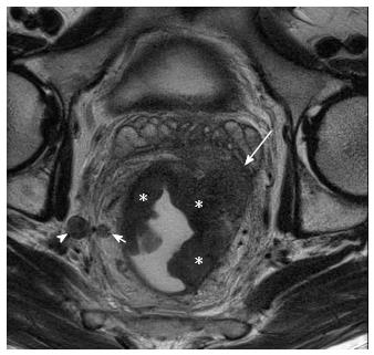
Figure 8 T3 tumor with lateral pelvic nodes.
Axial T2W magnetic resonance imaging shows a large right lateral pelvic wall node (arrowhead). Short arrow shows perirectal node. The primary rectal tumor (asterisk) is seen extending into left mesorectal fat upto the mesorectal fascia (long arrow).
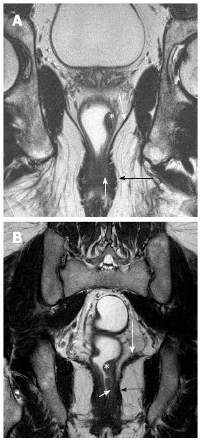
Figure 10 Coronal section T2 weighted magnetic resonance imaging to see level of tumor for planning surgery.
A and B show tumor (asterisk) along the left lateral wall, that reaches up to the internal sphincter (short white arrows) in B, but spares it in A. The uninvolved external sphincter (darkly hypointense) is shown by black arrows. The long white arrows in A and B shows the spared levator ani.
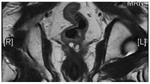
Figure 11 Low rectal cancer involving left levator.
- Citation: Saklani AP, Bae SU, Clayton A, Kim NK. Magnetic resonance imaging in rectal cancer: A surgeon’s perspective. World J Gastroenterol 2014; 20(8): 2030-2041
- URL: https://www.wjgnet.com/1007-9327/full/v20/i8/2030.htm
- DOI: https://dx.doi.org/10.3748/wjg.v20.i8.2030


