
















©2014 Baishideng Publishing Group Co.
World J Gastroenterol. Feb 28, 2014; 20(8): 2030-2041
Published online Feb 28, 2014. doi: 10.3748/wjg.v20.i8.2030
Published online Feb 28, 2014. doi: 10.3748/wjg.v20.i8.2030
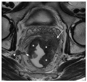
Figure 8 T3 tumor with lateral pelvic nodes.
Axial T2W magnetic resonance imaging shows a large right lateral pelvic wall node (arrowhead). Short arrow shows perirectal node. The primary rectal tumor (asterisk) is seen extending into left mesorectal fat upto the mesorectal fascia (long arrow).
- Citation: Saklani AP, Bae SU, Clayton A, Kim NK. Magnetic resonance imaging in rectal cancer: A surgeon’s perspective. World J Gastroenterol 2014; 20(8): 2030-2041
- URL: https://www.wjgnet.com/1007-9327/full/v20/i8/2030.htm
- DOI: https://dx.doi.org/10.3748/wjg.v20.i8.2030