Copyright
©2014 Baishideng Publishing Group Inc.
World J Gastroenterol. Dec 28, 2014; 20(48): 18503-18506
Published online Dec 28, 2014. doi: 10.3748/wjg.v20.i48.18503
Published online Dec 28, 2014. doi: 10.3748/wjg.v20.i48.18503
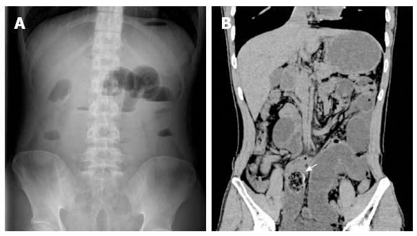
Figure 1 Radiologic findings.
A: Plain abdominal radiograph showed gas-fluid levels on left side of the abdomen; B: Computed tomography scan revealed a low intestinal obstruction (white arrow).
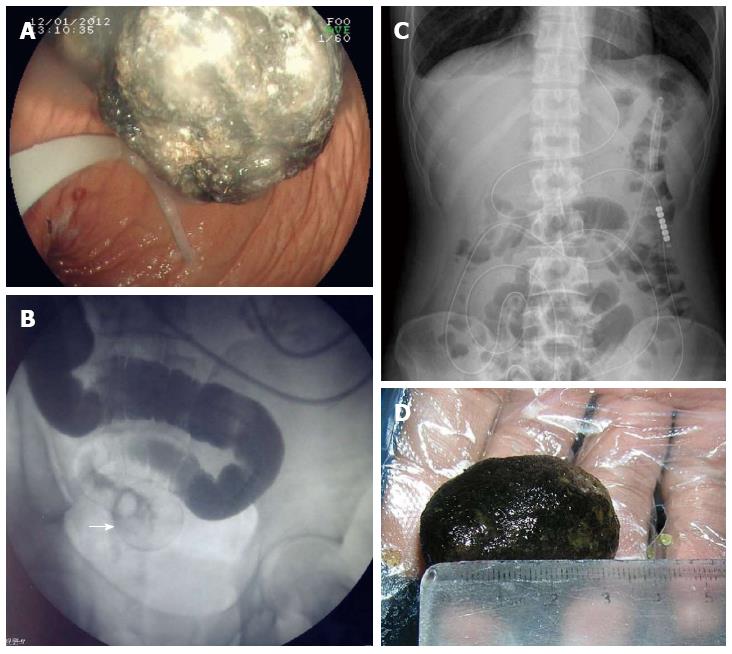
Figure 2 Treatment procedures.
A: A greenish, semisolid mass in the stomach observed on gastroscopy; B: Imaging revealed slight expansion of the intestine and dislodgement at a round point (white arrow); C: No air-fluid level was observed on plain abdominal radiograph two days later; D: Image of the discharged diospyrobezoar.
- Citation: Zheng YX, Prasoon P, Chen Y, Hu L, Chen L. ''Sandwich'' treatment for diospyrobezoar intestinal obstruction: A case report. World J Gastroenterol 2014; 20(48): 18503-18506
- URL: https://www.wjgnet.com/1007-9327/full/v20/i48/18503.htm
- DOI: https://dx.doi.org/10.3748/wjg.v20.i48.18503


