Copyright
©2014 Baishideng Publishing Group Inc.
World J Gastroenterol. Dec 28, 2014; 20(48): 18413-18419
Published online Dec 28, 2014. doi: 10.3748/wjg.v20.i48.18413
Published online Dec 28, 2014. doi: 10.3748/wjg.v20.i48.18413
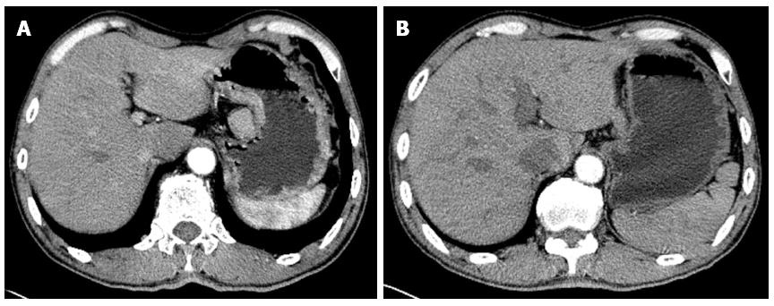
Figure 1 Comparison of computed tomography images before and after neoadjuvant chemotherapy.
A: The gastric wall in the gastric body exhibited irregular thickening, with the greatest thickness being 17 mm, and the enhancement was apparent. The gastric wall of the lesser curvature side showed an enlarged lymph node, with a size of about 32 mm × 23 mm, and its boundary with the stomach was still clear; B: Partial gastric wall of the gastric body thickened, showing a protruding mass, with a size of about 17 mm × 15 mm; this lesion showed heterogeneous enhancement. The outside wall of the stomach was smooth, the gap surrounding fat existed, and an enlarged lymph node could be seen at the lesser curvature side, with a size of about 17 mm × 11 mm.
Figure 2 Comparison of double contrast-enhanced ultrasonography images before and after neoadjuvant chemotherapy.
A: The gastric antrum, gastric angle, pylorus, partial gastric body and gastric wall thickened, involving the duodenal bulb, with the range of approximately 85 mm × 50 mm; the angiography showed fast-in-fast-out performance, part broke through the serosa, and multiple swelled lymph nodes could be seen near the gastric lesions, head of the pancreas and abdominal aorta, with the largest size being approximately 25 mm × 14 mm; B: The gastric antrum, gastric angle, pylorus, partial gastric body and gastric wall thickened, involving the duodenal bulb, with the range of approximately 60 mm × 35 mm; angiography showed fast-in-fast-out performance, the part broke through the serosa, multiple swelled lymph nodes could be seen near the gastric lesions, head of the pancreas and abdominal aorta, and the largest size was approximately 14 mm × 10 mm.
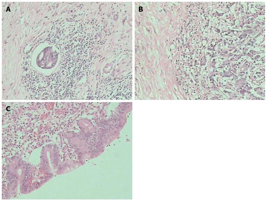
Figure 3 Comparison of pathological images before and after the neoadjuvant chemotherapy.
A: A small intravascular tumour thrombus could be seen in the middle of the tumour nest, and there were more fibrous tissues and scattered cancer cells around the tumour nest; B: More inflammatory cells could be seen surrounding the tumour tissues, with more fibrous tissues surrounding the inflammatory cells; C: There were many cancer cells with karyopyknosis and vacuole-like cells.
- Citation: Yu YJ, Sun WJ, Lu MD, Wang FH, Qi DS, Zhang Y, Li PH, Huang H, You T, Zheng ZQ. Efficacy of docetaxel combined with oxaliplatin and fluorouracil against stage III/IV gastric cancer. World J Gastroenterol 2014; 20(48): 18413-18419
- URL: https://www.wjgnet.com/1007-9327/full/v20/i48/18413.htm
- DOI: https://dx.doi.org/10.3748/wjg.v20.i48.18413


