Copyright
©2014 Baishideng Publishing Group Inc.
World J Gastroenterol. Dec 28, 2014; 20(48): 18296-18305
Published online Dec 28, 2014. doi: 10.3748/wjg.v20.i48.18296
Published online Dec 28, 2014. doi: 10.3748/wjg.v20.i48.18296
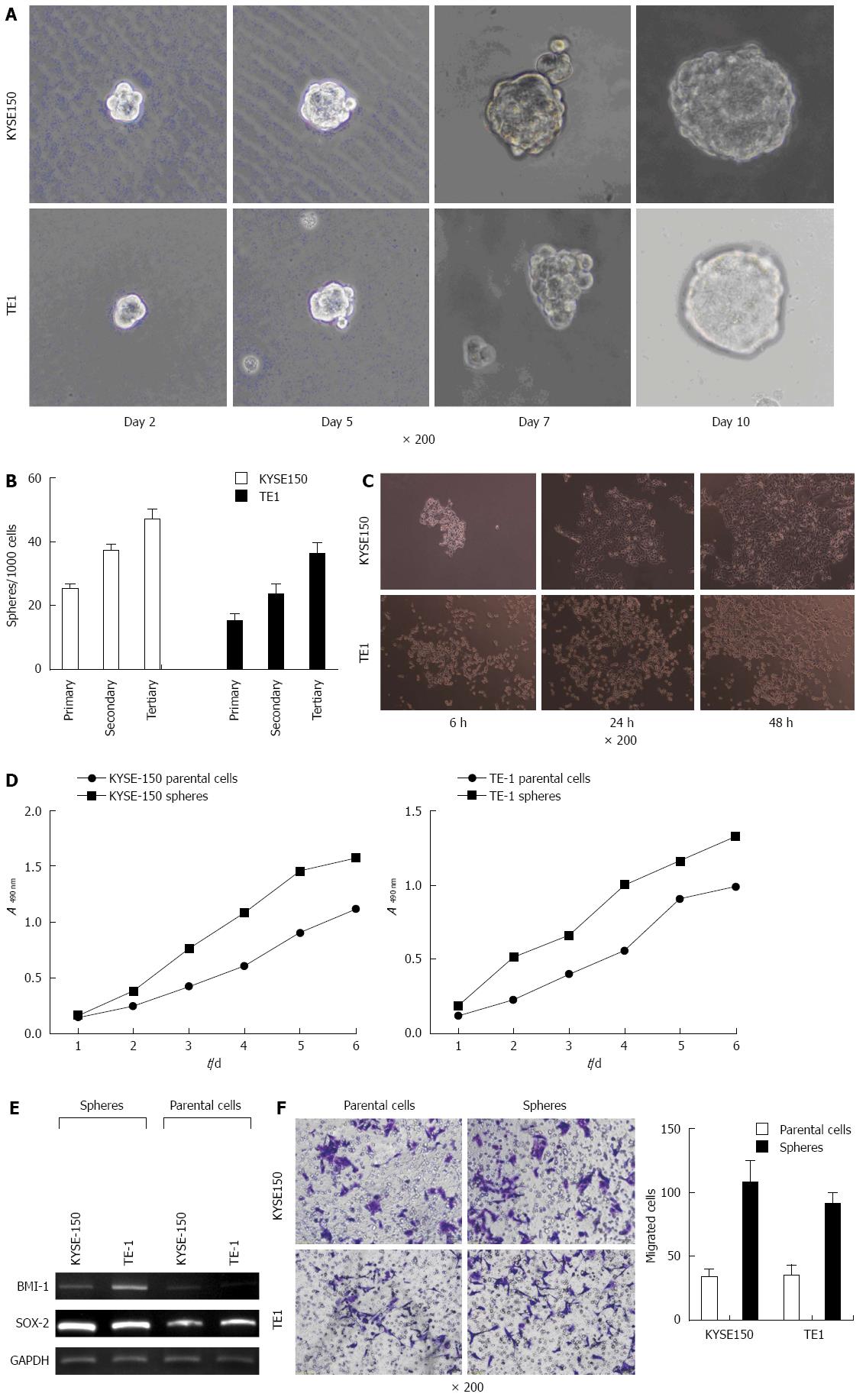
Figure 1 Differences in the biological characteristics of parent cells and spheres.
A: Cell spheres of different sizes suspended in the medium. The size of cell spheres gradually increased (× 200) with extended culture time; B: As the passage number increased, the rate of formation of cell spheres increased; C: Microscopic observation indicated that most cell spheres adhered after 6, 24 and 48 h; D: Cell growth was determined by the MTT assay. The multiplication capacity of KYSE150 and TE1 cell spheres was significantly higher than that of normal adherent cells; E: The expression of stem cell genes, BMI-1 and SOX-2, in cell spheres was increased; F: Transwell migration experiments demonstrating the invasion ability of KYSE150 and TE1 cell spheres in comparison with the invasion ability of parent cells.
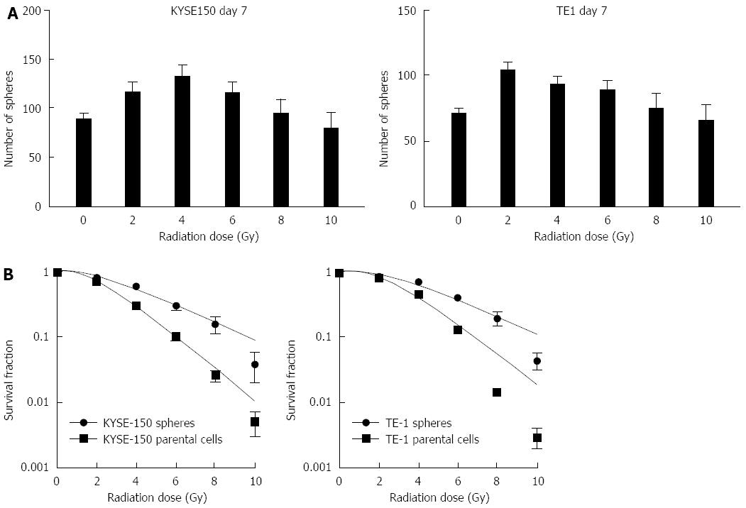
Figure 2 Rates of two types of cells generating stem-like spheres and differences in radiation sensitivity between stem-like spheres and parental cells.
A: Influence of irradiation dosage on cell sphere formation. Formation rates were increased at doses of 2, 4 and 6 Gy in the KYSE150 cell line and at doses of 2 and 4 Gy in the TE1 cell line (P < 0.05); B: The differences in sensitivity to radiation between cell spheres and adherent cells were compared in a clone formation assay. Cell spheres were also more radioresistant than parent cells (P < 0.05).
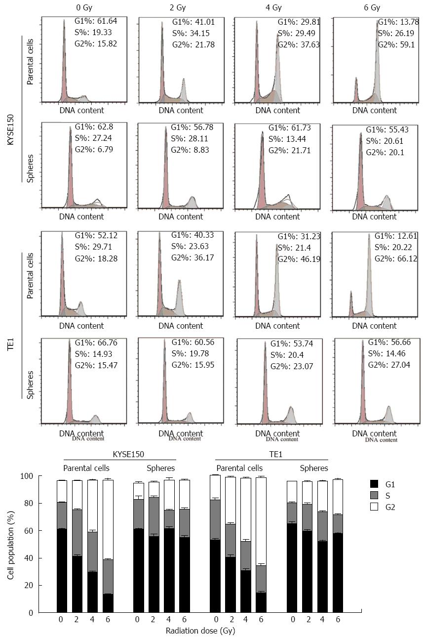
Figure 3 Influence of irradiation on cell cycle distribution changes.
KYSE150 and TE1 parent cells were clearly retarded at G2 phase, whereas the respective cell spheres were not.
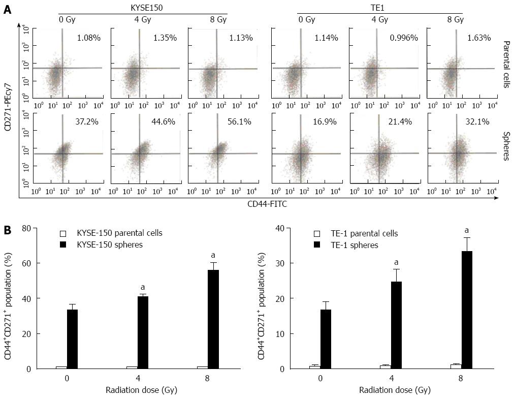
Figure 4 Proportion changes of CD44+CD271+ cells in parental cells and stem-like spheres with different doses of radiation.
A: Expression of CD44+, CD271+ and CD44+CD271+ were detected by flow cytometry; B: The differences in CD44+CD271+ cell proportion in parent cells and cell spheres under different irradiation doses were significant. In addition, the proportion of KYSE150 and TE1 cell spheres under irradiation doses of 4 and 8 Gy was significantly different from that of 0 Gy, aP < 0.05 vs control group.
- Citation: Wang JL, Yu JP, Sun ZQ, Sun SP. Radiobiological characteristics of cancer stem cells from esophageal cancer cell lines. World J Gastroenterol 2014; 20(48): 18296-18305
- URL: https://www.wjgnet.com/1007-9327/full/v20/i48/18296.htm
- DOI: https://dx.doi.org/10.3748/wjg.v20.i48.18296


