Copyright
©2014 Baishideng Publishing Group Inc.
World J Gastroenterol. Dec 21, 2014; 20(47): 18053-18056
Published online Dec 21, 2014. doi: 10.3748/wjg.v20.i47.18053
Published online Dec 21, 2014. doi: 10.3748/wjg.v20.i47.18053
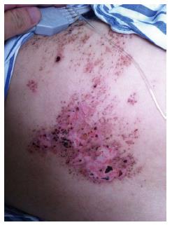
Figure 1 Presentation of characteristic rash.
Image showing the rash, which had begun to scab, on the patient’s right thoracodorsal area.
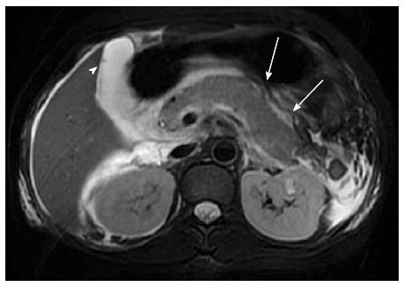
Figure 2 Magnetic resonance cholangiopancreatography.
Image showing a punctiform low signal intensity in the gallbladder (arrowhead) and peri-pancreatic exudation (arrows).
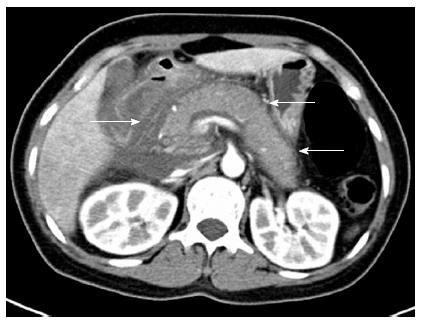
Figure 3 Contrast-enhanced computed tomography.
Image showing the swelling of the pancreas with peri-pancreatic exudation and liquid collection (arrows). Grade E acute pancreatitis was diagnosed based on the computed tomography severity index.
- Citation: Wang Z, Ye J, Han YH. Acute pancreatitis associated with herpes zoster: Case report and literature review. World J Gastroenterol 2014; 20(47): 18053-18056
- URL: https://www.wjgnet.com/1007-9327/full/v20/i47/18053.htm
- DOI: https://dx.doi.org/10.3748/wjg.v20.i47.18053


