Copyright
©2014 Baishideng Publishing Group Inc.
World J Gastroenterol. Dec 21, 2014; 20(47): 17924-17931
Published online Dec 21, 2014. doi: 10.3748/wjg.v20.i47.17924
Published online Dec 21, 2014. doi: 10.3748/wjg.v20.i47.17924
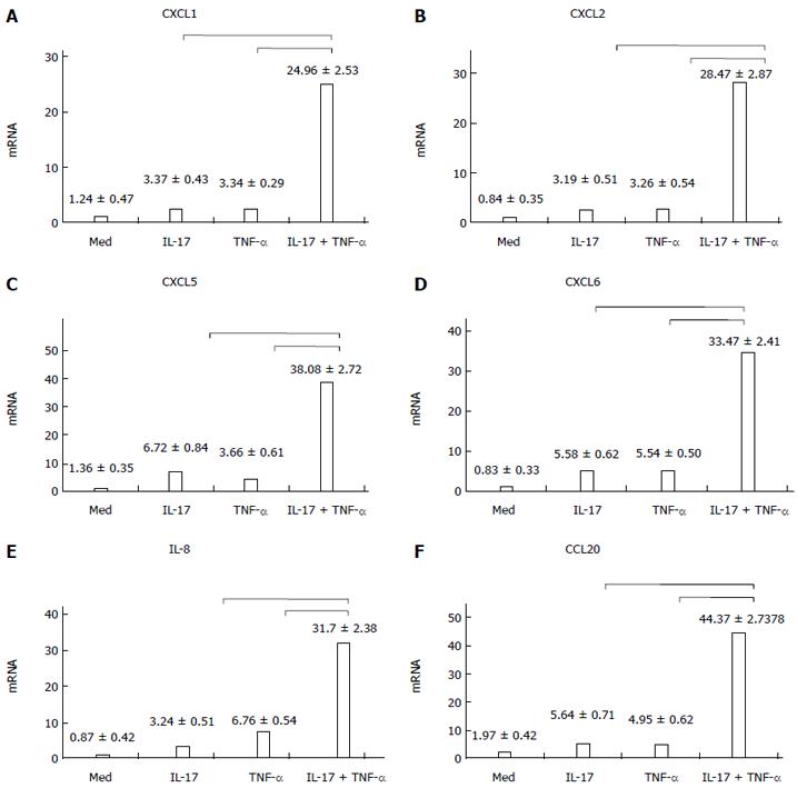
Figure 1 Chemokine expression when HT-29 cells were cultured with interleukin-17 and/or tumor necrosis factor-α.
When interleukin (IL)-17 and tumor necrosis factor (TNF)-α were used separately, the expression levels of CXCL1, CXCL2, CXCL5, CXCL6, IL-8 and CCL20 in HT-29 cells were comparatively low. However, when IL-17 and TNF-α were used together, the expression levels became strongly upregulated; A: IL-17 + TNF-αvs IL-17, t = 30.424, P = 0.000; IL-17 + TNF-αvs TNF-α, t = 30.438, P = 0.000; B: IL-17 + TNF-αvs IL-17, t = 35.273, P = 0.000; IL-17 + TNF-αvs TNF-α, t = 35.125, P = 0.000; C: IL-17 + TNF-αvs IL-17, t = 85.718, P = 0.000; IL-17 + TNF-αvs TNF-α, t = 94.199, P = 0.000; D: IL-17 + TNF-αvs IL-17, t = 50.165, P = 0.000; IL-17 + TNF-αvs TNF-α, t = 50.224, P = 0.000; E: IL-17 + TNF-αvs IL-17, t = 54.585, P = 0.000; IL-17 + TNF-αvs TNF-α, t = 44.036, P = 0.000; F: IL-17 + TNF-αvs IL-17, t = 56.272, P = 0.000; IL-17 + TNF-αvs TNF-α, t = 57.410, P = 0.000. IL-8: Interleukin-8.
Figure 2 p38 and extracellular signal-regulated kinase phosphorylation levels.
When interleukin (IL)-17 and tumor necrosis factor (TNF)-α were used separately, the phosphorylation levels of p38 and extracellular signal-regulated kinase (ERK) were very low. However, when IL-17 and TNF-α were combined together, the phosphorylation levels of p38 and ERK improved significantly, and the longer IL-17 and TNF-α were combined together, the stronger the phosphorylation became.
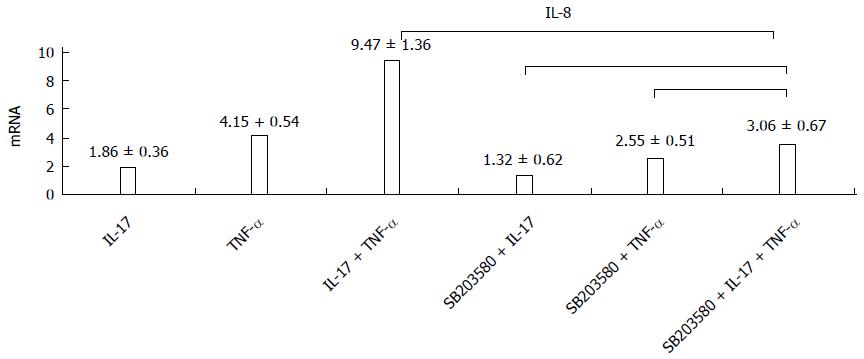
Figure 3 p38 inhibitor decreases the expression of interleukin-8.
When p38 inhibitor was used, the interleukin (IL)-8 expression levels declined in all three groups. IL-17 + TNF-αvs S + IL-17 + TNF-α, t = 103.701, P = 0.000; S + IL-17 + TNF-αvs S + TNF-α, t = - 4.131, P = 0.07; S + IL-17 + TNF-αvs S + IL-17, t = -11.509, P = 0.000. IL-8: Interleukin-8; TNF-α: Tumor necrosis factor-α; S: SB203580.
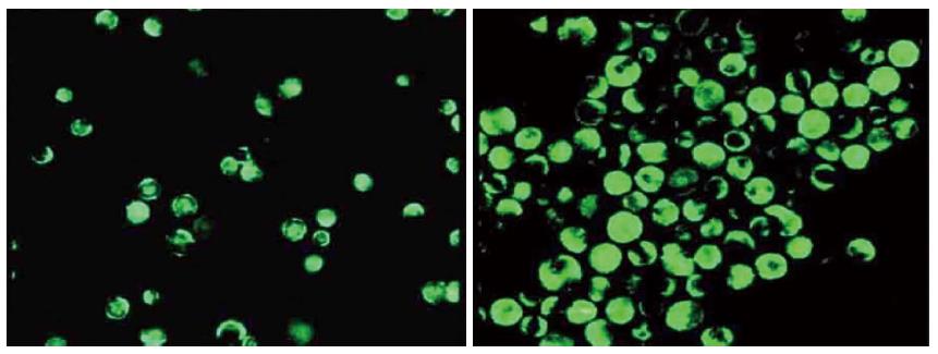
Figure 4 Act1 silenced HT-29 cells.
Compared with the control group (A), the expression level of Act1 was comparatively low in Act1 silenced cells (B).
Figure 5 Phosphorylation levels of p38 in Act1 silenced HT-29 cell line.
In the control group, interleukin (IL)-17 combined with tumor necrosis factor (TNF)-α could greatly improve the phosphorylation level of p38. In Act1 silenced cells, the phosphorylation level of p38 remained unchanged, when IL-17 and TNF-α were used together.
- Citation: Wang YL, Fang M, Wang XM, Liu WY, Zheng YJ, Wu XB, Tao R. Proinflammatory effects and molecular mechanisms of interleukin-17 in intestinal epithelial cell line HT-29. World J Gastroenterol 2014; 20(47): 17924-17931
- URL: https://www.wjgnet.com/1007-9327/full/v20/i47/17924.htm
- DOI: https://dx.doi.org/10.3748/wjg.v20.i47.17924


