Copyright
©2014 Baishideng Publishing Group Inc.
World J Gastroenterol. Dec 21, 2014; 20(47): 17746-17755
Published online Dec 21, 2014. doi: 10.3748/wjg.v20.i47.17746
Published online Dec 21, 2014. doi: 10.3748/wjg.v20.i47.17746
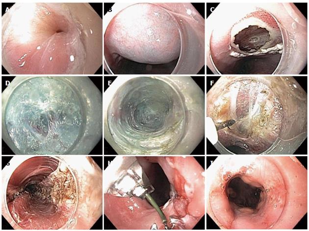
Figure 1 Per-oral endoscopic myotomy technique.
A: Prior to per-oral endoscopic myotomy (POEM), there is evidence of a tightly puckered lower esophageal sphincter (LES); B: Submucosal injection is performed with saline stained with indigo carmine; C: Mucosotomy is performed along the right anterior wall of the esophagus; D: Submucosal dissection is performed with hybrid knife; E: Submucosal tunnel is extended into the gastric cardia; F: Myotomy is intiated 2 cm below site of mucosotomy; G: Final full thickness myotomy is seen as endoscope is withdrawn from the submucosal tunnel; H: Mucosotomy is closed with an endoscopic suturing device; I: After POEM, the LES appears patulous.
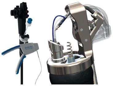
Figure 2 OverStitch endoscopic suturing system (Courtesy of Apollo Endosurgery, Austin Texas).
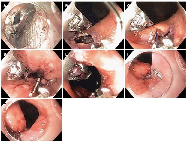
Figure 3 Closure of gastroesophageal junction mucosal perforation with endoscopic suturing device.
A: Inadvertent mucosal perforation at the gastroesophageal junction (GEJ) seen within the submucosal tunnel; B: Inadvertent mucosal perforation at the GEJ seen endoluminally; C: Endoscopic suture-initial “bite”; D: Suture closure; E: Cinch T-tag is deployed; F: Endoscopic suturing achieved secure closure of the perforation; G: Patulous lower esophageal sphincter.
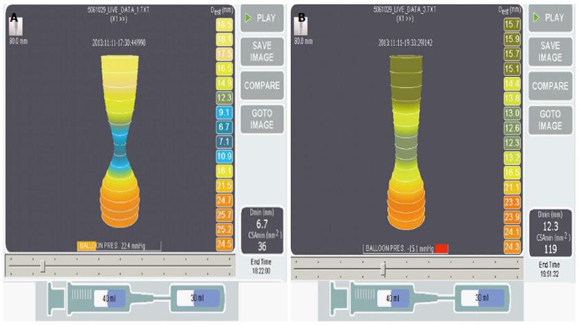
Figure 4 EndoFLIP Images before and after per-oral endoscopic myotomy.
Seventy-seven years old man with achalasia for 4 years, prior Botox × 1, esophageal diameter of 5 cm, non-sigmoid, underwent per-oral endoscopic myotomy (POEM), 8 cm posterior myotomy. A: EndoFLIP performed immediately prior to POEM; B: immediately after POEM at 30 mL balloon volume shows an excellent response with increase of the minimal diameter at the gastroesophageal junction (GEJ) (Dmin) from 6.7 to 12.3 cm and increase in cross-sectional diameter at the GEJ (cross-sectional area) from 36 to 119 mm2.
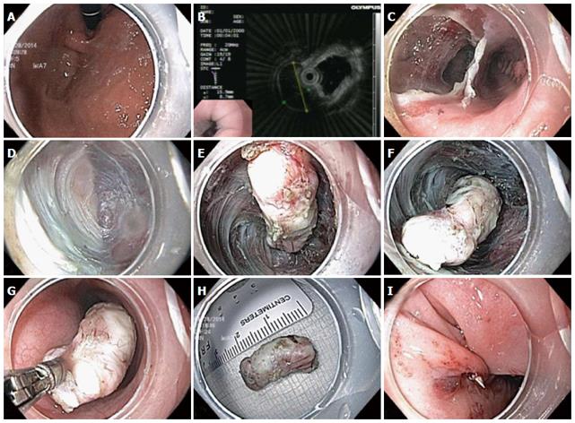
Figure 5 Submucosal tunneling endoscopic resection technique.
A: Gastroesophageal junction lesion seen on retroflexion during endoscopy; B: Endoscopic ultrasound probe demonstrates hypoechoic muscularis propria based lesion; C: Creation of submucosal tunnel parallel to esophageal lumen; D: Endoscopic submucosal dissection (ESD) with submucosal tunnel; E-F: Freeing of submucosal lesion via ESD; G: Removal of submucosal lesion from tunnel with biopsy forceps; H: 2.5 cm leiomyoma; I: Endoscopic sutured closure of mucosal entrance to tunnel.
- Citation: Friedel D, Modayil R, Stavropoulos SN. Per-oral endoscopic myotomy: Major advance in achalasia treatment and in endoscopic surgery. World J Gastroenterol 2014; 20(47): 17746-17755
- URL: https://www.wjgnet.com/1007-9327/full/v20/i47/17746.htm
- DOI: https://dx.doi.org/10.3748/wjg.v20.i47.17746


