Copyright
©2014 Baishideng Publishing Group Inc.
World J Gastroenterol. Dec 14, 2014; 20(46): 17686-17689
Published online Dec 14, 2014. doi: 10.3748/wjg.v20.i46.17686
Published online Dec 14, 2014. doi: 10.3748/wjg.v20.i46.17686
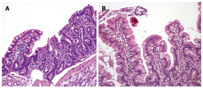
Figure 1 Duodenal biopsy specimens before (A) and 6 mo after (B) gluten-free diet in our patient with Selective IgM deficiency and seronegative celiac disease.
A subtotal villous atrophy with an almost flat mucosa is evident before, and complete villous restoration after the dietary therapy (HE: × 200).
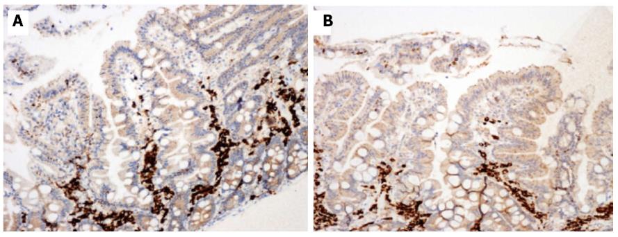
Figure 2 Duodenal biopsy specimens before (A) and 12 mo after (B) gluten-free diet in our patient with Selective IgM deficiency and seronegative celiac disease.
Immunohistochemistry showed a mild reduction of CD 79a immature lymphocytes. Positive cells are stained brown and counterstained blue (× 200).
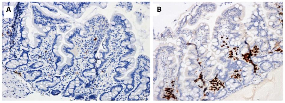
Figure 3 Duodenal biopsy specimens before (A) and 12 mo after (B) gluten-free diet in our patient with Selective IgM deficiency and seronegative celiac disease.
Immunohistochemistry showed scarse MUM1 plasma cells before the diet and a marked positivity 12 mo later. Positive cells are stained brown and counterstained blue (× 200).
- Citation: Montenegro L, Piscitelli D, Giorgio F, Covelli C, Fiore MG, Losurdo G, Iannone A, Ierardi E, Di Leo A, Principi M. Reversal of IgM deficiency following a gluten-free diet in seronegative celiac disease. World J Gastroenterol 2014; 20(46): 17686-17689
- URL: https://www.wjgnet.com/1007-9327/full/v20/i46/17686.htm
- DOI: https://dx.doi.org/10.3748/wjg.v20.i46.17686


