Copyright
©2014 Baishideng Publishing Group Inc.
World J Gastroenterol. Dec 14, 2014; 20(46): 17434-17438
Published online Dec 14, 2014. doi: 10.3748/wjg.v20.i46.17434
Published online Dec 14, 2014. doi: 10.3748/wjg.v20.i46.17434
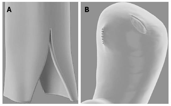
Figure 1 Oesophageal flaps and “sleeve joint”.
A: A longitudinal cut on the distal oesophageal about 2-3 cm in length; B: A stoma in the anterior wall of the stomach forming a “sleeve joint”, which was 2 to 3 mm longer than the oesophageal diameter.
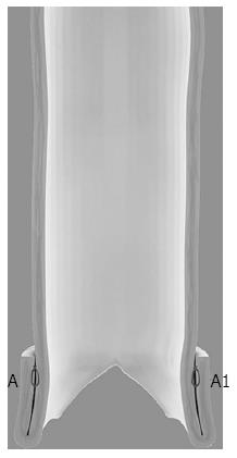
Figure 2 Oesophageal flap valvuloplasty.
The seromuscular layer of the interrupted absorbable monofilament suture was used to suture the 1 cm-long fold around the remnant oesophageal flaps in the AA1 site forming modified oesophageal flaps.
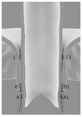
Figure 3 Wrapping suturing of the stomach wall around the oesophagus.
The seromuscular layer of the oesophagus and the gastric wall was sutured in the BB1 site and CC1 site. The BB1 site was located just above the end of the oesophageal flaps and the distance between BB1 and CC1 should be 2 cm.
- Citation: Dai JG, Liu QX, Den XF, Min JX. Oesophageal flap valvuloplasty and wrapping suturing prevent gastrooesophageal reflux disease in dogs after oesophageal anastomosis. World J Gastroenterol 2014; 20(46): 17434-17438
- URL: https://www.wjgnet.com/1007-9327/full/v20/i46/17434.htm
- DOI: https://dx.doi.org/10.3748/wjg.v20.i46.17434


