Copyright
©2014 Baishideng Publishing Group Inc.
World J Gastroenterol. Dec 14, 2014; 20(46): 17399-17406
Published online Dec 14, 2014. doi: 10.3748/wjg.v20.i46.17399
Published online Dec 14, 2014. doi: 10.3748/wjg.v20.i46.17399
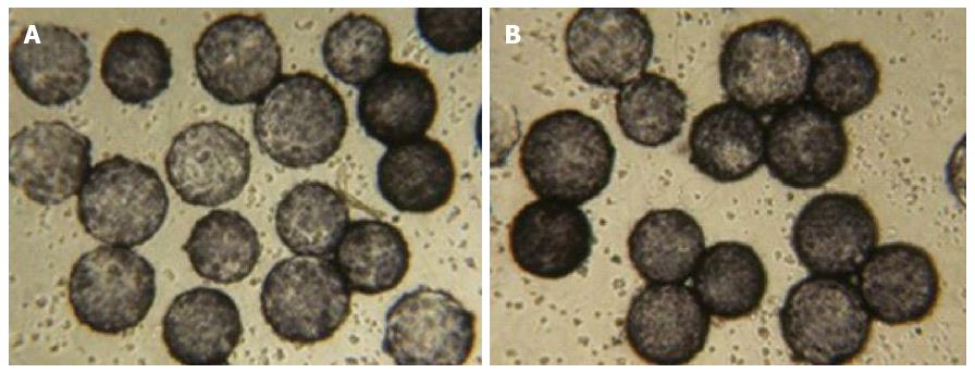
Figure 1 Cell viability.
Trypan blue staining in Chinese human liver cells seeded in a Rotary Cell Culture System A: Before hybrid bioartificial liver support system treatment (cell viability: 95%-99%); and B: after treatment (cell viability: 85%-89%); magnification × 100.
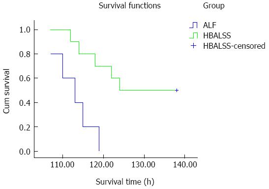
Figure 2 Survival curves.
In the hybrid bioartificial liver support system (HBALSS) treatment group, five cynomolgus monkeys survived and the remaining five died. The survival time was 128 ± 3 h. In control groups, all cynomolgus monkeys died and the survival time was 112 ± 2 h. ALF: Acute liver failure.
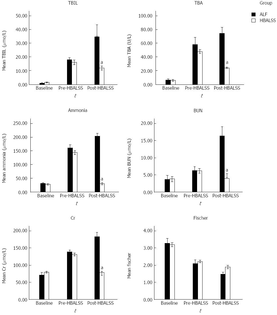
Figure 3 Serum biochemistry.
Before and after D-galactosamine injection, serum concentrations of total bilirubin (TBiL), total bile acid (TBA), ammonia, blood urea nitrogen (BUN) and creatinine (Cr) were significantly increased, whereas Fischer indices were significantly reduced. Six hours after treatment in the acute liver failure (ALF) group, levels of TBiL, TBA, ammonia, BUN, and Cr remained elevated, while the Fischer indices remained decreased. Additionally, in the hybrid bioartificial liver support system (HBALSS) group, levels of TBiL, TBA, ammonia, BUN and Cr decreased significantly. aP < 0.05 vs control group.
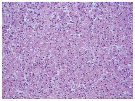
Figure 4 Light microscopy of a liver biopsy.
After the passing of the animal subjects, liver tissues were prepared and stained for hematoxylin and eosin. Hepatocytes showed diffused swelling, were sinusoidal, and became narrow after pressure. The cytoplasm of hepatocytes appeared loose, and there were signs of vacuolar degeneration, nuclear fragmentation and dissolution. Part of the portal area showed a small amount of neutrophils and lymphocytes (× 200).
- Citation: Zhang Z, Zhao YC, Cheng Y, Jian GD, Pan MX, Gao Y. Hybrid bioartificial liver support in cynomolgus monkeys with D-galactosamine-induced acute liver failure. World J Gastroenterol 2014; 20(46): 17399-17406
- URL: https://www.wjgnet.com/1007-9327/full/v20/i46/17399.htm
- DOI: https://dx.doi.org/10.3748/wjg.v20.i46.17399


