Copyright
©2014 Baishideng Publishing Group Inc.
World J Gastroenterol. Dec 7, 2014; 20(45): 17179-17184
Published online Dec 7, 2014. doi: 10.3748/wjg.v20.i45.17179
Published online Dec 7, 2014. doi: 10.3748/wjg.v20.i45.17179
Figure 1 Two overlapping stent-in-stent technique.
A: Computed tomography angiography (CTA) showing a thrombus generated inside the false lumen, an ulcer-like niche intruding into the false lumen (indicated by the arrow); B: DSA demonstrating isolated superior mesenteric artery dissection (ISMAD) and narrowing of native lumen (arrow); C: Post two bare stent (a balloon-expandable and a self-expandable bare) deployment, repeat angiograph showing complete exclusion of the true lumen preserving patency of the superior mesenteric artery; D: Follow-up CTA at 5 mo, demonstrating disappearance of the false lumen and patency of true lumen (arrow).
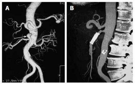
Figure 2 Single bare stent technique.
A: An isolated superior mesenteric artery dissection detected by computed tomography angiography (indicated by the arrow); B: Patent true lumen and the disappearance of the false lumen at 3 mo after single bare stenting.
- Citation: Lv PH, Zhang XC, Wang LF, Chen ZL, Shi HB. Management of isolated superior mesenteric artery dissection. World J Gastroenterol 2014; 20(45): 17179-17184
- URL: https://www.wjgnet.com/1007-9327/full/v20/i45/17179.htm
- DOI: https://dx.doi.org/10.3748/wjg.v20.i45.17179


