Copyright
©2014 Baishideng Publishing Group Inc.
World J Gastroenterol. Dec 7, 2014; 20(45): 17163-17170
Published online Dec 7, 2014. doi: 10.3748/wjg.v20.i45.17163
Published online Dec 7, 2014. doi: 10.3748/wjg.v20.i45.17163
Figure 1 Circulating tumor cells captured and cultured on the membrane filter (A), cultured in vitro (A, B, C, D) with visualized nucleoli and nucleus counterstained with DAPI.
The circulating tumor cells (CTCs) cultured in vitro may grow through the membrane via the ability of changing the cell shape (C, D).
Figure 2 Circulating tumor cells captured and cultured on the membrane filter, incubated with CK-18-FITC antibody, nucleus was counterstained by DAPI (A, B).
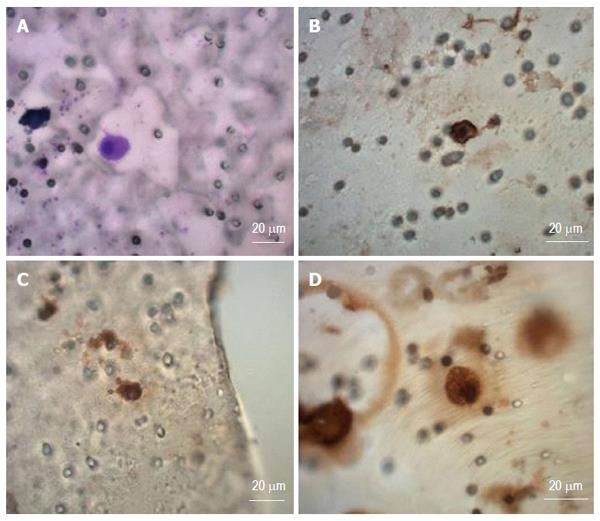
Figure 3 Circulating tumor cells captured on the separating membrane shown after different immunohistochemistry stainig proving the gastrointestinal origine of the captured cells.
A: May-Gruenwald stain; B: CEA-antibody staining; C: CK7-antibody staining; D: Vimentin-antibody staining.
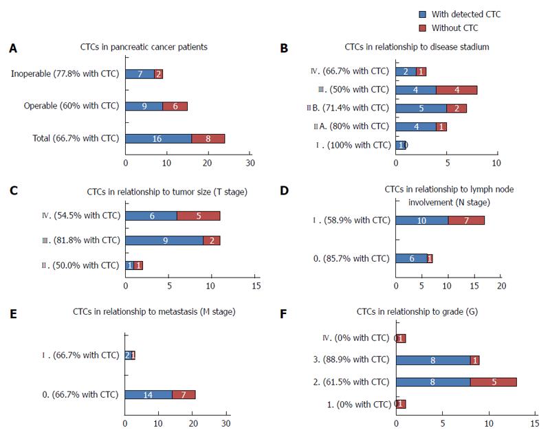
Figure 4 Circulating tumor cells positivity reported for patient subgroups.
A: Circulating tumor cells (CTCs) in PaC patients, reflecting operability of the primary tumor; B: CTCs in PaC patients in different disease stages; C: CTCs in PaC patients, grouped according the tumor size; D: CTCs in PaC patients, grouped according the lymph node involvement; E: CTCs in PaC patients grouped according the metastasis; F: CTCs in PaC patients, grouped according to tumor grade.
- Citation: Bobek V, Gurlich R, Eliasova P, Kolostova K. Circulating tumor cells in pancreatic cancer patients: Enrichment and cultivation. World J Gastroenterol 2014; 20(45): 17163-17170
- URL: https://www.wjgnet.com/1007-9327/full/v20/i45/17163.htm
- DOI: https://dx.doi.org/10.3748/wjg.v20.i45.17163


