Copyright
©2014 Baishideng Publishing Group Inc.
World J Gastroenterol. Nov 28, 2014; 20(44): 16550-16558
Published online Nov 28, 2014. doi: 10.3748/wjg.v20.i44.16550
Published online Nov 28, 2014. doi: 10.3748/wjg.v20.i44.16550
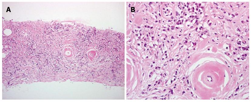
Figure 1 Histological findings with core needle biopsy: Fibrosis and inflammatory reaction with dense infiltration of abundant lymphocytes and plasma cells.
A: Low magnification; B: High magnification.
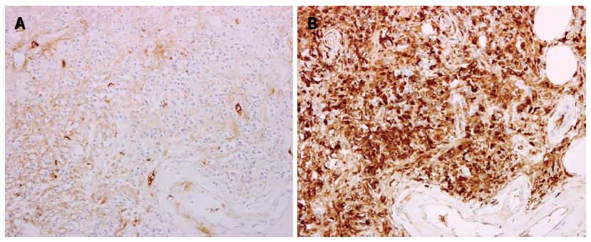
Figure 2 Immunopathological findings: infiltrating cells represent lymphocytes and IgG4-positive plasma cells.
A: IgG4 staining; B: IgM staining.
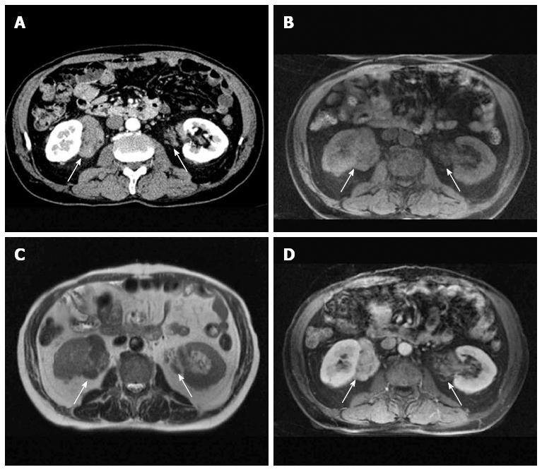
Figure 3 Localized pseudotumors (arrows) in a 67-year-old man with IgG4-related retroperitoneal fibrosis and autoimmune pancreatitis.
A: Contrast-enhanced CT; B: T1-weighted; C: T2-weighted; D: Contrast-enhanced MRI.
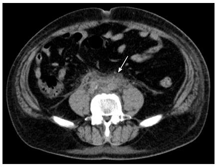
Figure 4 Pseudotumor spread in the retroperitoneum surrounding the abdominal aorta and vena cava (arrow).
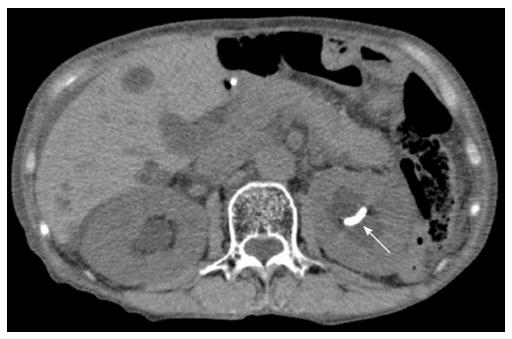
Figure 5 Although a ureteral stent was correctly placed into the left renal pelvis (arrow), ipsilateral hydronephrosis did not improve in a 79-year-old woman with IgG4-related retroperitoneal fibrosis.
- Citation: Hara N, Kawaguchi M, Takeda K, Zen Y. Retroperitoneal disorders associated with IgG4-related autoimmune pancreatitis. World J Gastroenterol 2014; 20(44): 16550-16558
- URL: https://www.wjgnet.com/1007-9327/full/v20/i44/16550.htm
- DOI: https://dx.doi.org/10.3748/wjg.v20.i44.16550


