Copyright
©2014 Baishideng Publishing Group Inc.
World J Gastroenterol. Nov 21, 2014; 20(43): 16359-16363
Published online Nov 21, 2014. doi: 10.3748/wjg.v20.i43.16359
Published online Nov 21, 2014. doi: 10.3748/wjg.v20.i43.16359
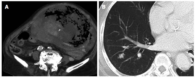
Figure 1 Preoperative computed tomography.
A: Abdominal computed tomography (CT) shows a giant tumor with central necrosis, extending from the epigastrium to the pelvic cavity; B: Chest CT shows multiple lung metastases.
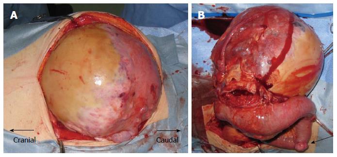
Figure 2 Intraoperative images.
A: On laparotomy, a giant, flexible tumor occupies the peritoneal cavity; B: The tumor is adherent to the small intestine and appears hemorrhagic. Multiple small tumors are recognized at other sites of the small intestine (arrow) and abdominal wall.
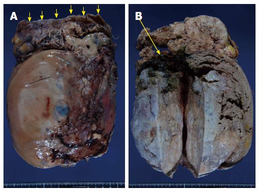
Figure 3 Resected specimen.
A: The resected tumor is solid, weighing 3490 g and measuring 24 cm × 17.5 cm × 18 cm. Continuity is seen between the tumor and small intestine (arrows show small intestine); B: The inside of the tumor appears necrotic. Furthermore, a hole (3 cm in diameter) is present between the tumor and small intestine, and a large amount of coagulated blood is present in the small intestine (arrow).
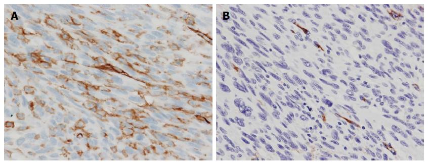
Figure 4 Histopathological examination.
A: Positive staining for CD31; B: Positive staining for factor VIII-related antigen.
- Citation: Takahashi M, Ohara M, Kimura N, Domen H, Yamabuki T, Komuro K, Tsuchikawa T, Hirano S, Iwashiro N. Giant primary angiosarcoma of the small intestine showing severe sepsis. World J Gastroenterol 2014; 20(43): 16359-16363
- URL: https://www.wjgnet.com/1007-9327/full/v20/i43/16359.htm
- DOI: https://dx.doi.org/10.3748/wjg.v20.i43.16359


