Copyright
©2014 Baishideng Publishing Group Inc.
World J Gastroenterol. Nov 21, 2014; 20(43): 16349-16354
Published online Nov 21, 2014. doi: 10.3748/wjg.v20.i43.16349
Published online Nov 21, 2014. doi: 10.3748/wjg.v20.i43.16349
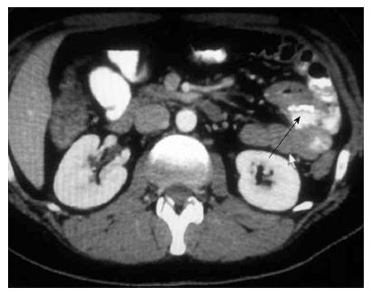
Figure 1 Jejuno-jejunal anastomosis on the left side of abdomen (arrow).
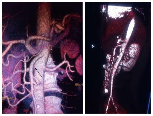
Figure 2 Major intensity projections computed tomography reconstructions can show the absence of loops behind the superior mesentery artery (arrow).
Figure 3 Major intensity projections computed tomography reconstructions show the Mesenteric fan deployed on the left side of abdomen (arrow).
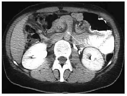
Figure 4 “Hurricane eye” computed tomography sign of internal hernia (arrow).
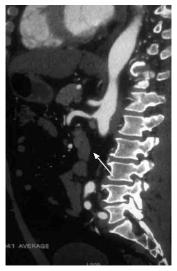
Figure 5 Small-bowel behind superior mesentery artery at computed tomography slice (arrow).
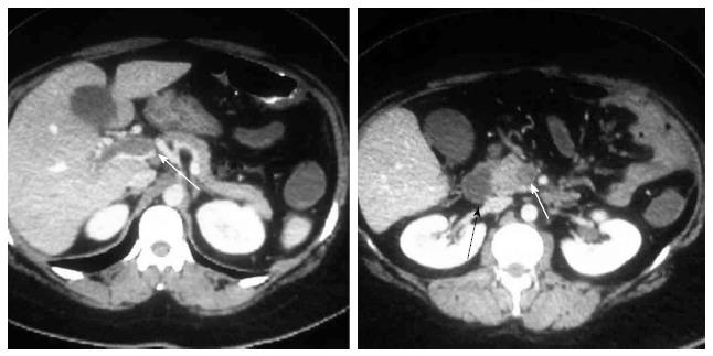
Figure 6 Portal and superior mesenteric thrombosis (arrow).
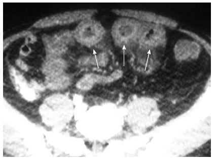
Figure 7 Computed tomography signs of “Clustered loops” (arrow).
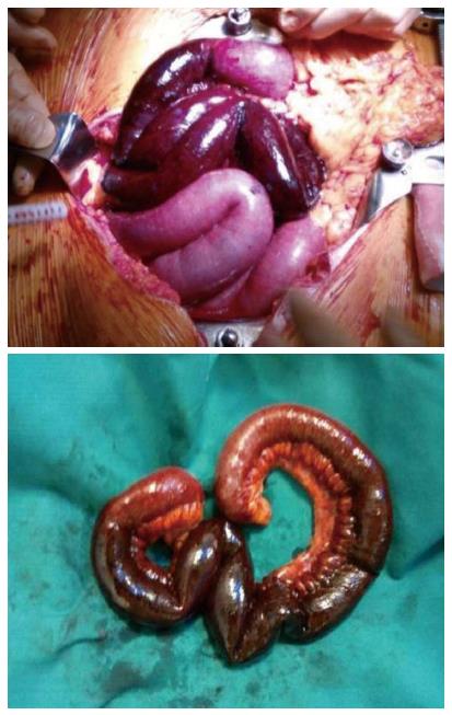
Figure 8 Small bowel infarction.
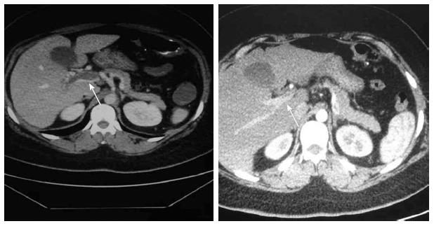
Figure 9 Comparison of computed tomography before and 2 mo after surgery shows the Portal and Mesenteric recanalization after the anticoagulant therapy (arrow).
- Citation: Fabozzi M, Brachet Contul R, Millo P, Allieta R. Intestinal infarction by internal hernia in Petersen’s space after laparoscopic gastric bypass. World J Gastroenterol 2014; 20(43): 16349-16354
- URL: https://www.wjgnet.com/1007-9327/full/v20/i43/16349.htm
- DOI: https://dx.doi.org/10.3748/wjg.v20.i43.16349


