Copyright
©2014 Baishideng Publishing Group Inc.
World J Gastroenterol. Nov 21, 2014; 20(43): 16268-16274
Published online Nov 21, 2014. doi: 10.3748/wjg.v20.i43.16268
Published online Nov 21, 2014. doi: 10.3748/wjg.v20.i43.16268
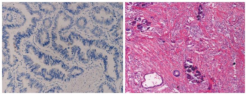
Figure 1 Insulin-like growth factor receptor-1 immunostaining of rectal cancer (× 200) and histopathological response to radiation (× 100) (a radiosensitivitive sample).
Negative cytoplasmic immunostaining of rectal cancer cells was seen on the pre-radiation biopsy specimen (left panel); correspondingly, good histopathological response to radiation therapy was seen on the post-operative sample (right panel): most of the entire lesion was replaced by fibrosis and necrosis, relic tumor glandular tubes and calcifications were seen.
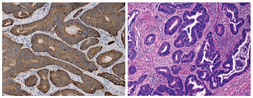
Figure 2 Insulin-like growth factor receptor-1 immunostaining of rectal cancer (× 200) and histopathological response to radiation (× 100) (a radioresistant sample).
Strong cytoplasmic immunostaining of rectal cancer cells was seen on the pre-radiation biopsy specimen (left panel), correspondingly, poor histopathological response to radiation therapy was seen on the post-operative sample (right panel): most of the tumor remained, and little fibrosis was seen among the tumor cells.
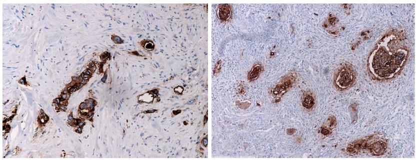
Figure 3 Insulin-like growth factor receptor-1 immunostaining in the post-radiation rectal specimens.
Regardless of radiosensitivity, strong cytoplasmic immunostaining (left panel) was often seen in the post-radiation remaining cancer cells, fibrosis was seen around the remaining tumor glandular tubes (right panel).
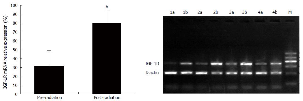
Figure 4 Insulin-like growth factor receptor-1 mRNA expression by reverse transcription polymerase chain reaction in the paired pre-radiation and post-radiation samples.
bP < 0.01 vs the paired pre-radiation specimens, the post-operative specimens showed a significantly increased insulin-like growth factor receptor-1 (IGF-1R) mRNA expression. M: Marker; 1-4a: Pre-radiation samples; 1-4b: Post-radiation samples.
- Citation: Wu XY, Wu ZF, Cao QH, Chen C, Chen ZW, Xu Z, Li WS, Liu FK, Yao XQ, Li G. Insulin-like growth factor receptor-1 overexpression is associated with poor response of rectal cancers to radiotherapy. World J Gastroenterol 2014; 20(43): 16268-16274
- URL: https://www.wjgnet.com/1007-9327/full/v20/i43/16268.htm
- DOI: https://dx.doi.org/10.3748/wjg.v20.i43.16268


