Copyright
©2014 Baishideng Publishing Group Inc.
World J Gastroenterol. Nov 21, 2014; 20(43): 16159-16166
Published online Nov 21, 2014. doi: 10.3748/wjg.v20.i43.16159
Published online Nov 21, 2014. doi: 10.3748/wjg.v20.i43.16159
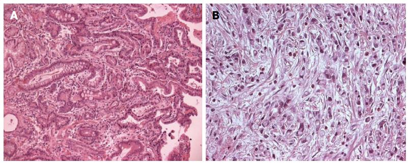
Figure 1 Intestinal-type adenocarcinoma.
Intestinal metaplastic epithelium is adjacent to the carcinoma (A), diffuse-type carcinoma composed of signet-ring cells showing foamy cytoplasm and an eccentrically located nucleus (B).
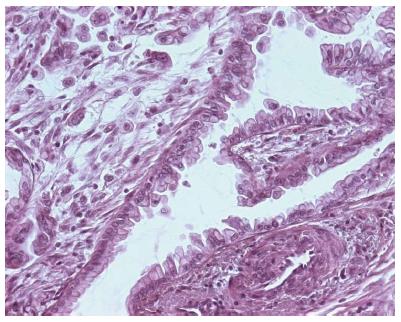
Figure 2 Gastric-type adenocarcinoma showing a papillary growth pattern admixed with a poorly differentiated component.
Neoplastic glands are lined by cuboidal to tall columnar cells showing clear mucinous cytoplasm and basally oriented enlarged nuclei.
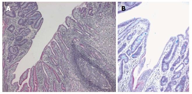
Figure 3 Gastric adenoma of intestinal type (A), dysplastic epithelium shows mild architectural changes with little branching or irregularity (left, note prominent lymphoid follicle in the adjacent non-neoplastic mucosa (right), alcian bleu- periodic acid Schiff; A few small goblet cells are scattered among columnar cells showing elongated nuclei (B) (Alcian blue- periodic acid Schiff).
- Citation: Rigoli L, Caruso RA. Mitochondrial DNA alterations in the progression of gastric carcinomas: Unexplored issues and future research needs. World J Gastroenterol 2014; 20(43): 16159-16166
- URL: https://www.wjgnet.com/1007-9327/full/v20/i43/16159.htm
- DOI: https://dx.doi.org/10.3748/wjg.v20.i43.16159


