Copyright
©2014 Baishideng Publishing Group Inc.
World J Gastroenterol. Nov 21, 2014; 20(43): 16020-16028
Published online Nov 21, 2014. doi: 10.3748/wjg.v20.i43.16020
Published online Nov 21, 2014. doi: 10.3748/wjg.v20.i43.16020
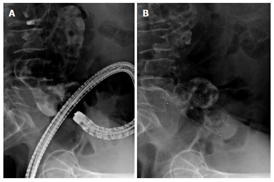
Figure 1 Self-expandable metallic stent placement for acute left-side malignant obstruction.
A: Fluoroscopy showed a malignant stricture with 3 cm length at a rectosigmoid junction. A guide wire was passed through the stricture; B: A 8 cm uncovered stent was successfully inserted and deployed.
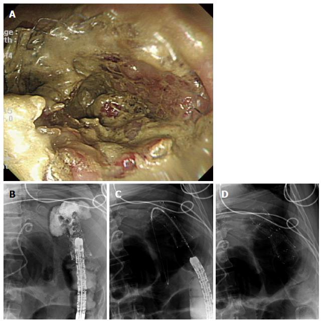
Figure 2 Stent in-stent placement for re-obstruction.
A: Colonoscopy showed a tumor in-growth in a previously inserted uncovered stent at splenic flexus; B: Fluoroscopy showed a significant narrowing of stent due to the tumor in-growth; C: A guide wire and a stent were inserted sequentially through the previously inserted stent; D: A 10 cm uncovered stent was successfully deployed without complications.
- Citation: Hong SP, Kim TI. Colorectal stenting: An advanced approach to malignant colorectal obstruction. World J Gastroenterol 2014; 20(43): 16020-16028
- URL: https://www.wjgnet.com/1007-9327/full/v20/i43/16020.htm
- DOI: https://dx.doi.org/10.3748/wjg.v20.i43.16020


