Copyright
©2014 Baishideng Publishing Group Inc.
World J Gastroenterol. Nov 14, 2014; 20(42): 15780-15786
Published online Nov 14, 2014. doi: 10.3748/wjg.v20.i42.15780
Published online Nov 14, 2014. doi: 10.3748/wjg.v20.i42.15780
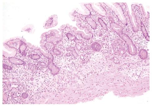
Figure 1 Gastric antrum mucosa (hematoxylin-eosin staining) with chronic atrophic gastritis and moderate-to-intense inflammatory infiltrate.
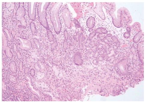
Figure 2 Gastric corpus mucosa: Loss of oxyntic cells and the presence of pseudo-pyloric metaplasia.
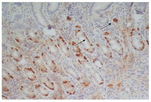
Figure 3 Chromogranin A staining of gastric corpus mucosa from a patient with autoimmune gastritis.
Note the linear and nodular (arrows) hyperplasia of endocrine cells.
- Citation: Gonçalves C, Oliveira ME, Palha AM, Ferrão A, Morais A, Lopes AI. Autoimmune gastritis presenting as iron deficiency anemia in childhood. World J Gastroenterol 2014; 20(42): 15780-15786
- URL: https://www.wjgnet.com/1007-9327/full/v20/i42/15780.htm
- DOI: https://dx.doi.org/10.3748/wjg.v20.i42.15780


