Copyright
©2014 Baishideng Publishing Group Inc.
World J Gastroenterol. Nov 7, 2014; 20(41): 15125-15134
Published online Nov 7, 2014. doi: 10.3748/wjg.v20.i41.15125
Published online Nov 7, 2014. doi: 10.3748/wjg.v20.i41.15125
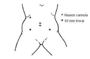
Figure 1 Schematic illustration of port placement for laparoscopic proctectomy.
Hasson cannula is placed first via a vertical infra-umbilical incision. The left lower quadrant port is not always used and is reserved for cases in which it is needed to facilitate splenic flexure mobilization and retraction at the time of pelvic dissection. It is usually needed in obese patients and in women with an enlarged uterus.
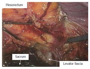
Figure 2 View of the pelvic dissection at the time of laparoscopic proctectomy.
Total mesorectal excision was carried out to the level of the levator muscles and the mesorectum is reflected superiorly. This quality of exposure is rarely seen in open total mesorectal excision.
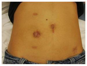
Figure 3 Cosmetic outcome after a laparoscopic assisted low anterior resection with a diverting loop ileostomy for rectal cancer, followed by a takedown of ileostomy.
The specimen was extracted through a periumbilical incision which, especially in thin persons, can be very limited.
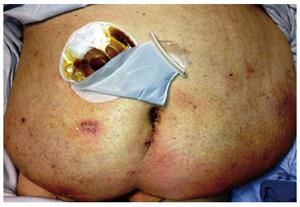
Figure 4 Laparoscopic assisted low anterior resection can be safely performed in morbid obese patients.
This is a 49 year old gentleman with a body mass index of 41 kg/m2. The picture was taken at the same hospital stay in which the patient underwent surgery.
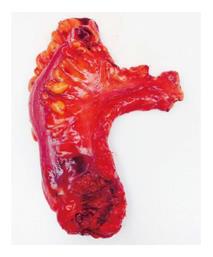
Figure 5 Specimen resected in a laparoscopic proctectomy.
Notice the high ligation of the inferior mesenteric artery and the shiny surface of the mesorectum, which reflects its intactness. The laparoscopic approach facilitates visualization and hence precise dissection and achievement of an intact mesorectum.
- Citation: Shussman N, Wexner SD. Current status of laparoscopy for the treatment of rectal cancer. World J Gastroenterol 2014; 20(41): 15125-15134
- URL: https://www.wjgnet.com/1007-9327/full/v20/i41/15125.htm
- DOI: https://dx.doi.org/10.3748/wjg.v20.i41.15125


