Copyright
©2014 Baishideng Publishing Group Inc.
World J Gastroenterol. Nov 7, 2014; 20(41): 15007-15017
Published online Nov 7, 2014. doi: 10.3748/wjg.v20.i41.15007
Published online Nov 7, 2014. doi: 10.3748/wjg.v20.i41.15007
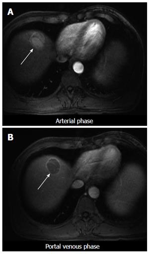
Figure 1 Magnetic resonance imaging of hepatocellular carcinoma in segment 8.
Arterial enhancement (A, arrow), washout and pseudocapsule (B, arrow).
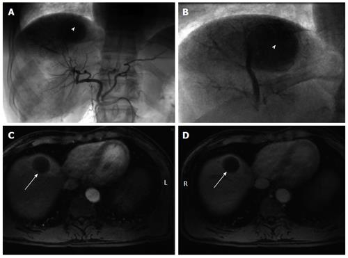
Figure 2 Treatment of hepatocellular carcinoma with transarterial chemoembolization.
A, B: Angiographic images demonstrate arterial blush of hepatocellular carcinoma (HCC) (white arrowhead) with a proper hepatic arterial injection. Coned down view shows tumor blush and stasis in the segmental arteries supplying the HCC following transarterial chemoembolization (B); C, D: Follow up magnetic resonance imaging 4 wk after treatment shows no residual arterial enhancement in the treated area (white arrows) compatible with a complete response per modified response evaluation criteria in solid tumors.
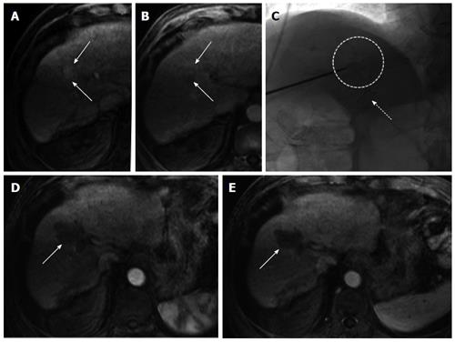
Figure 3 Pretreatment of patients on transplant list (other modalities).
A and B: Arterial enhancing hepatocellular carcinoma lesion in segment 5/8 (arrows) that shows subtle washout and possible pseudocapsule on delayed post-contrast imaging. This tumor was treated successfully using percutaneous microwave ablation; C: The tip of the microwave ablation probe positioned within the lesion (white dotted circle). Note the presence of a transjugular intrahepatic portosystemic shunt catheter (white dotted arrow) which helped direct placement of the probe on fluoroscopic images; D, E: Follow up magnetic resonance imaging 4 wk after ablation shows large ablation cavity covering the region of the previously seen tumor (arrows). There is no residual arterial enhancement suggesting complete tumor necrosis.
- Citation: Khan AS, Fowler KJ, Chapman WC. Current surgical treatment strategies for hepatocellular carcinoma in North America. World J Gastroenterol 2014; 20(41): 15007-15017
- URL: https://www.wjgnet.com/1007-9327/full/v20/i41/15007.htm
- DOI: https://dx.doi.org/10.3748/wjg.v20.i41.15007


