Copyright
©2014 Baishideng Publishing Group Co.
World J Gastroenterol. Jan 28, 2014; 20(4): 1088-1094
Published online Jan 28, 2014. doi: 10.3748/wjg.v20.i4.1088
Published online Jan 28, 2014. doi: 10.3748/wjg.v20.i4.1088
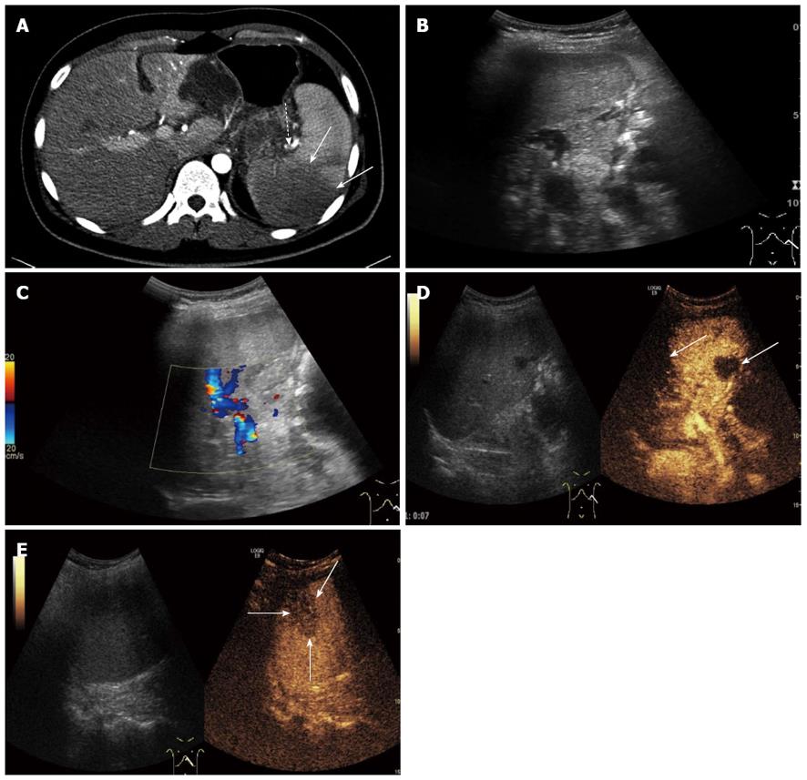
Figure 1 A 22-year-old man with splenic artery embolism.
A: Enhanced-contrast computed tomography showing splenic artery embolism (dotted arrow), and the lesion of splenic infarction (solid arrow); B, C: Gray scale ultrasonography and color Doppler ultrasonography of the spleen showed no obvious lesion of splenic infarction; D: Some regions with no enhancement appeared in the splenic arterial phase (solid arrow) on contrast enhanced ultrasound (CEUS); E: A wedge-shaped splenic infarction with no enhancement was displayed in the splenic venous phase (solid arrow) on CEUS.
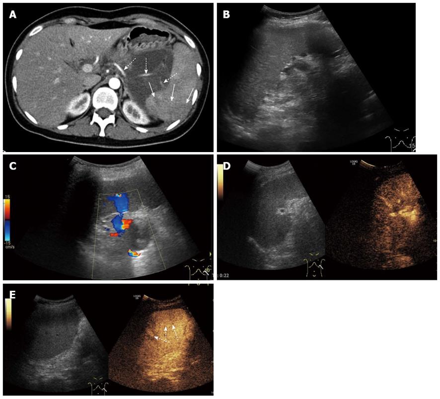
Figure 2 A 26-year-old woman with splenic arterial stenosis.
A: Enhanced-contrast computed tomography showing splenic arterial stenosis (dotted arrow) and a focal splenic parenchyma region showed low enhancement (solid arrow); B, C: Gray scale ultrasonography and color Doppler ultrasonography showed no obvious lesions in the spleen; D: No obvious lesion was displayed in the splenic arterial phase on contrast enhanced ultrasound (CEUS); E: Some regions of focal dysperfusion of the splenic parenchyma showed low enhancement in the splenic venous phase (dotted arrow).
- Citation: Cai DM, Parajuly SS, Ling WW, Li YZ, Luo Y. Diagnostic value of contrast enhanced ultrasound for splenic artery complications following acute pancreatitis. World J Gastroenterol 2014; 20(4): 1088-1094
- URL: https://www.wjgnet.com/1007-9327/full/v20/i4/1088.htm
- DOI: https://dx.doi.org/10.3748/wjg.v20.i4.1088


