Copyright
©2014 Baishideng Publishing Group Co.
World J Gastroenterol. Jan 28, 2014; 20(4): 1079-1087
Published online Jan 28, 2014. doi: 10.3748/wjg.v20.i4.1079
Published online Jan 28, 2014. doi: 10.3748/wjg.v20.i4.1079
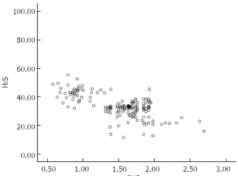
Figure 1 Negative correlation between H2S plasma levels and portal vein diameters in patients with portal hypertension.
r = -0.478, P < 0.05.
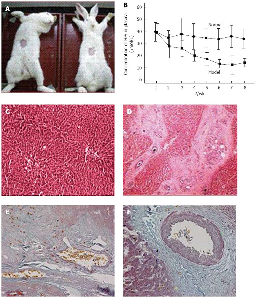
Figure 2 Liver hematoxylin and eosin and Masson’s trichrome staining of rabbits in portal hypertension model group and control group.
A: Rabbit portal hypertension model; B: Relationship between rabbit portal hypertension progression and H2S concentration; C: Normal rabbit liver tissue [hematoxylin and eosin (HE) staining]; D: Schistosomiasis portal hypertension (SPH) rabbit liver sample with concentric arrangement of fibrous larval nodules and fibrous connective tissue in the portal area (HE staining); E and F: Masson’s trichrome staining: collagen fibers are stained blue-green; muscle fibers and cellulose are stained red; and nuclei are stained blue to black; E: Normal liver tissue; F: SPH liver tissue with large amount of collagen fibers (arrow). All histological images are shown with × 40 magnification.
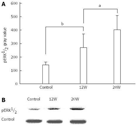
Figure 3 Western blotting analysis of phosphorylated extracellular signal-regulated kinase 1/2 expression levels in schistosomiasis portal hypertension and control rabbit portal vein-tunica media dissections.
aP < 0.05,bP < 0.001.
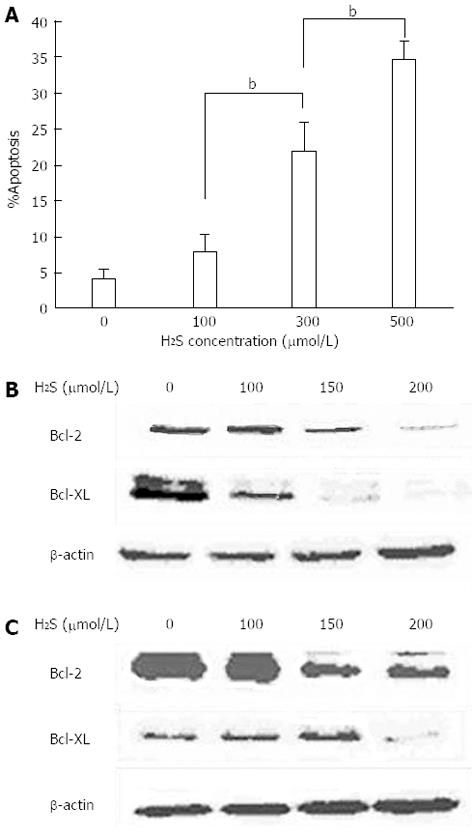
Figure 4 (A) Smooth muscle cell apoptosis under varying H2S concentrations, observed by flow cytometry, (B) Protein levels of the apoptotic factors Bcl-2 and Bcl-XL in primary rabbit portal vein smooth muscle cells and (C) in primary human portal vein smooth muscle cells.
bP < 0.001.
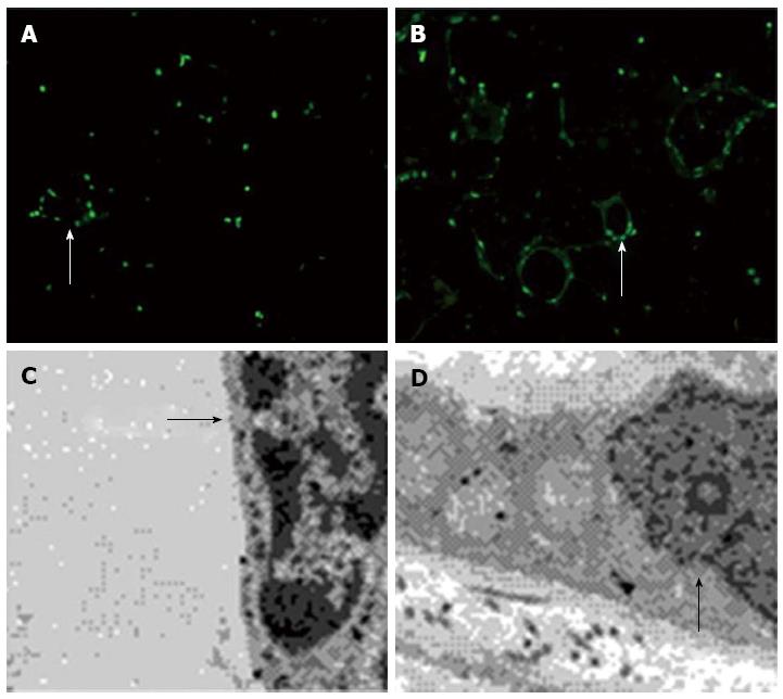
Figure 5 Effect of H2S concentrations on portal vein smooth muscle cell apoptosis rates.
A: Immunofluorescence assays showed significant apoptosis in normal rabbit omentum vascular smooth muscle cells under normal H2S (50 μmol/L) concentration; B: There was significantly less apoptosis in the cells without H2S (magnification × 40); C: Under an electron microscope, with normal H2S concentrations, rupturing of the nuclear membrane and condensation of the nucleoplasm was observed, indicating irreversible cell death; D: In cells without H2S substitution, the mitochondria appeared intact and the structural integrity was maintained.
- Citation: Wang C, Han J, Xiao L, Jin CE, Li DJ, Yang Z. Role of hydrogen sulfide in portal hypertension and esophagogastric junction vascular disease. World J Gastroenterol 2014; 20(4): 1079-1087
- URL: https://www.wjgnet.com/1007-9327/full/v20/i4/1079.htm
- DOI: https://dx.doi.org/10.3748/wjg.v20.i4.1079


