Copyright
©2014 Baishideng Publishing Group Inc.
World J Gastroenterol. Oct 21, 2014; 20(39): 14420-14429
Published online Oct 21, 2014. doi: 10.3748/wjg.v20.i39.14420
Published online Oct 21, 2014. doi: 10.3748/wjg.v20.i39.14420
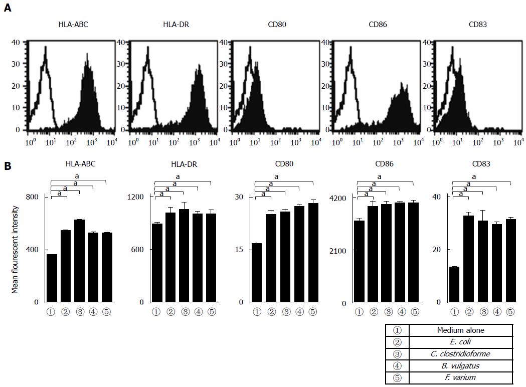
Figure 1 Phenotypic characterization of dendritic cells.
A: Human monocyte-derived dendritic cells (MoDCs) were analyzed using flow cytometry for expression of the indicated antigens (n = 5). Unfilled histogram profiles indicate the isotype control, and solid histograms indicate the specific antibodies; B: Analysis of the mean fluorescence intensity of the indicated molecules expressed in MoDCs stimulated with commensal bacteria (n = 5). Unstimulated MoDCs were used as a control. The results are expressed as the mean ± SD. aP < 0.05.
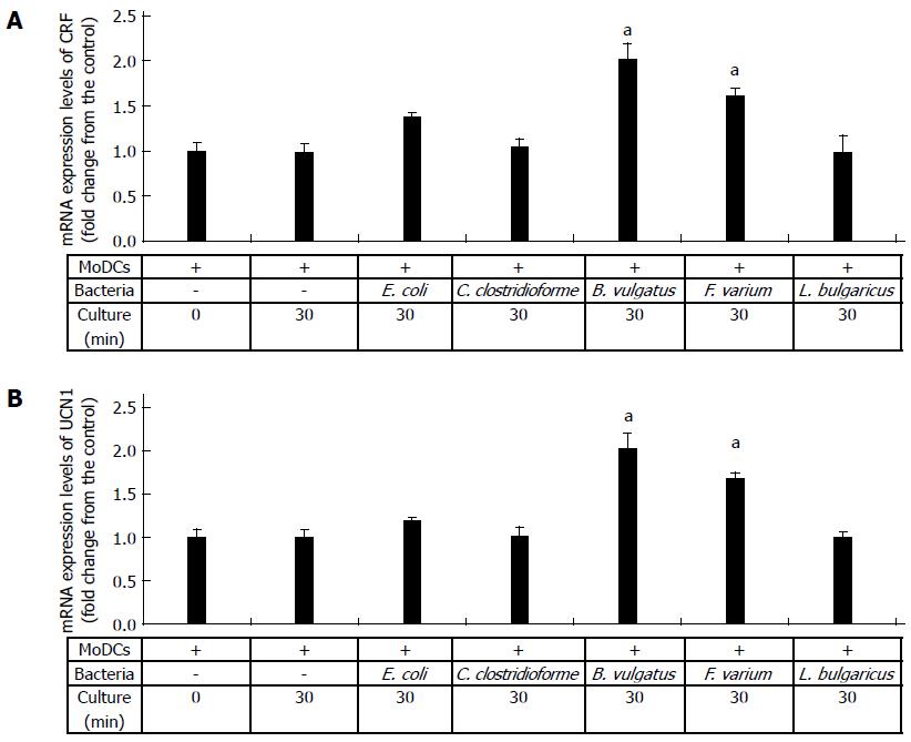
Figure 2 Detection of corticotropin-releasing factor and urocortin 1 mRNA in dendritic cells stimulated with commensal bacteria.
Human monocyte-derived dendritic cells (MoDCs) were stimulated with Escherichia coli (E. coli), Clostridium clostridioforme (C. clostridioforme), Bacteroides vulgatus (B. vulgatus), Fusobacterium varium (F. varium), and Lactobacillus delbrueckii subsp. bulgaricus (L. bulgaricus) for 30 min, and A: corticotropin-releasing factor (CRF) and B: urocortin 1 (UCN1) mRNA were analyzed. Human MoDCs were cultured alone for 0 and 30 min and used as controls. CT values reflect the polymerase chain reaction cycle number at which the amount of amplified target reached a fixed threshold. The results are expressed as the mean ± SD from 3 repeated experiments. aP < 0.05 vs other groups.
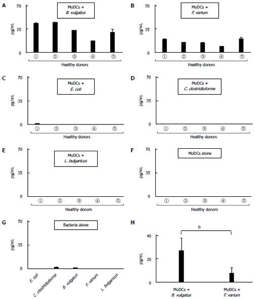
Figure 3 Production of corticotropin-releasing factor in dendritic cells stimulated with commensal bacteria.
Human monocyte-derived DCs (MoDCs) from 5 healthy donors (①, ②, ③, ④, and ⑤) were stimulated with A: Bacteroides vulgatus (B. vulgatus); B: Fusobacterium varium (F. varium); C: Escherichia coli (E. coli); D: Clostridium clostridioforme (C. clostridioforme); E: Lactobacillus delbrueckii subsp. bulgaricus (L. bulgaricus) for 24 h, after which the production of corticotropin-releasing factor (CRF) was analyzed. Supernatants from F: human MoDCs alone and G: commensal bacteria alone were used as controls; H: The production of CRF in human MoDCs stimulated with B. vulgatus was compared with that in human MoDCs stimulated with F. varium. The results are expressed as the mean ± SD from 5 healthy donors. bP < 0.01, MoDCs + B. vulgatus vs MoDCs s + F. varium.
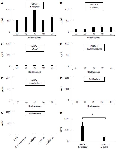
Figure 4 Production of urocortin 1 in dendritic cells stimulated with commensal bacteria.
Human monocyte-derived DCs (MoDCs) from 5 healthy donors (①, ②, ③, ④, and ⑤) were stimulated with A: Bacteroides vulgatus (B. vulgatus); B: Fusobacterium varium (F. varium); C: Escherichia coli (E. coli); D: Clostridium clostridioforme (C. clostridioforme); E: Lactobacillus delbrueckii subsp. bulgaricus (L. bulgaricus) for 24 h, after which the production of urocortin 1 (UCN1) was analyzed. Supernatants from F: human MoDCs alone and G: commensal bacteria alone were used as controls; H: The production of UCN1 in human MoDCs stimulated with B. vulgatus was compared with that in human MoDCs stimulated with F. varium. The results are expressed as the mean ± SD from 5 healthy donors. bP < 0.01, MoDCs + B. vulgatus vs MoDCs s + F. varium.
- Citation: Koido S, Ohkusa T, Kan S, Takakura K, Saito K, Komita H, Ito Z, Kobayashi H, Takami S, Uchiyama K, Arakawa H, Ito M, Okamoto M, Kajihara M, Homma S, Tajiri H. Production of corticotropin-releasing factor and urocortin from human monocyte-derived dendritic cells is stimulated by commensal bacteria in intestine. World J Gastroenterol 2014; 20(39): 14420-14429
- URL: https://www.wjgnet.com/1007-9327/full/v20/i39/14420.htm
- DOI: https://dx.doi.org/10.3748/wjg.v20.i39.14420


