Copyright
©2014 Baishideng Publishing Group Inc.
World J Gastroenterol. Oct 14, 2014; 20(38): 13820-13832
Published online Oct 14, 2014. doi: 10.3748/wjg.v20.i38.13820
Published online Oct 14, 2014. doi: 10.3748/wjg.v20.i38.13820
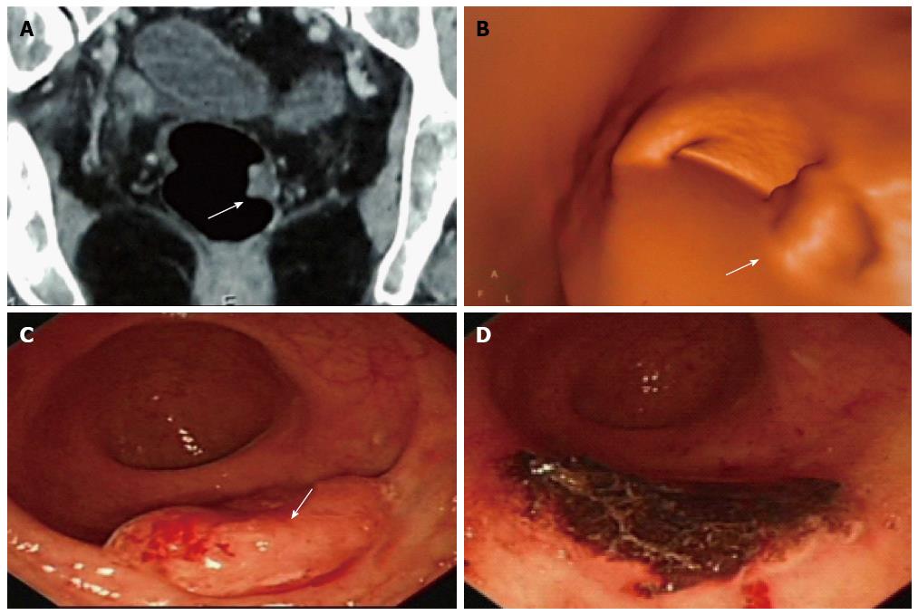
Figure 1 Combination use of computed tomography, computed tomography colonography and conventional colonoscopy in large bowel examination and management.
A 62-year-old woman complained of blood stool for several days and was detected with multi-polyps in colorectal cancer screening. Virtual and conventional colonoscopy all had positive results (single white arrow) and pathology after removal under conventional colonoscopy (CC) proved that the most suspected polyp had turned to be moderately differentiated adenocarcinoma. A: Lesion revealed by cross-sectional computed tomography (CT); B: Same lesion by CT colonography (CTC); C: By CC; D: CC after removal of the cancer. The case illustrated the different roles of CT, CTC and CC, as well as the combination use of these modalities. The invasiveness of CC might explain injury and swelling of the lesion, and the role of CC in polypectomy is demonstrated.
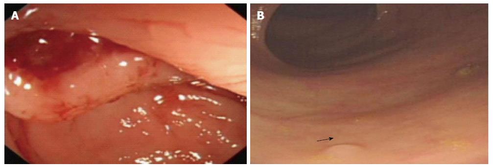
Figure 2 Examples of risk and merit of conventional colonoscopy.
A: A 51-year-old woman was found with perforation of the colon during conventional colonoscopy; B: A 68-year-old man was detected with multi-polyps with the smallest diameter of 2 mm (arrow) in one routine colorectal cancer screening.
Figure 3 Standard low-dose positron emission tomography/computed tomography showing the colon and rectum.
- Citation: He Q, Rao T, Guan YS. Virtual gastrointestinal colonoscopy in combination with large bowel endoscopy: Clinical application. World J Gastroenterol 2014; 20(38): 13820-13832
- URL: https://www.wjgnet.com/1007-9327/full/v20/i38/13820.htm
- DOI: https://dx.doi.org/10.3748/wjg.v20.i38.13820


