Copyright
©2014 Baishideng Publishing Group Inc.
World J Gastroenterol. Oct 14, 2014; 20(38): 13648-13657
Published online Oct 14, 2014. doi: 10.3748/wjg.v20.i38.13648
Published online Oct 14, 2014. doi: 10.3748/wjg.v20.i38.13648
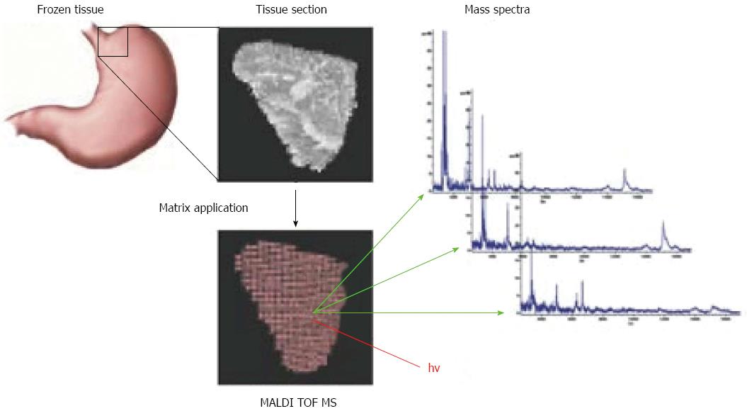
Figure 1 Principle of matrix-assisted laser desorption-ionization imaging mass spectrometry.
A section is cut from frozen tissue and prepared for mass spectrometry by coating with matrix solution. Energy for desorption and ionization is supplied by a pulsed laser beam. For each point in the user-defined measurement grid, a mass spectrum is generated by MALDI-TOF MS[79]. Copyright © 2009, Rights Managed by Nature Publishing Group. MALDI: Matrix-assisted laser desorption-ionization; TOF: Time-of-flight; MS: Mass spectrometry.
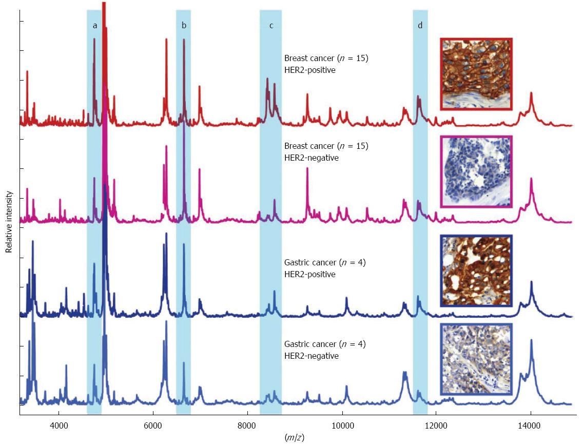
Figure 2 Matrix-assisted laser desorption-ionization imaging mass spectrometric profiles of breast and gastric cancer tissues.
Human epidermal growth factor receptor-2 (HER2)-status can be identified on a proteomic level across different cancer types suggesting that HER2 overexpression may constitute a unique molecular event independent of the tumor site[65]. Reprinted with permission from [65]. Copyright © 2010 American Chemical Society.
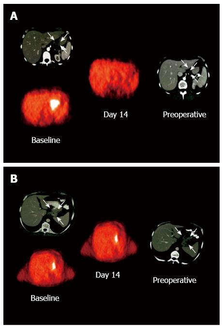
Figure 3 Positron emission tomography with the glucose analog fluorine-18 fluorodeoxyglucose studies in patients with clinically responding and nonresponding tumors[75].
A: In the responding tumor, fluorodeoxyglucose (FDG) uptake decreases to background level 14 d after initiation of chemotherapy; B: In contrast, FDG uptake is almost unchanged for the nonresponding tumor. Copyright © 2003 American Society of Clinical Oncology.
- Citation: Aichler M, Luber B, Lordick F, Walch A. Proteomic and metabolic prediction of response to therapy in gastric cancer. World J Gastroenterol 2014; 20(38): 13648-13657
- URL: https://www.wjgnet.com/1007-9327/full/v20/i38/13648.htm
- DOI: https://dx.doi.org/10.3748/wjg.v20.i38.13648


