Copyright
©2014 Baishideng Publishing Group Inc.
World J Gastroenterol. Oct 7, 2014; 20(37): 13556-13562
Published online Oct 7, 2014. doi: 10.3748/wjg.v20.i37.13556
Published online Oct 7, 2014. doi: 10.3748/wjg.v20.i37.13556
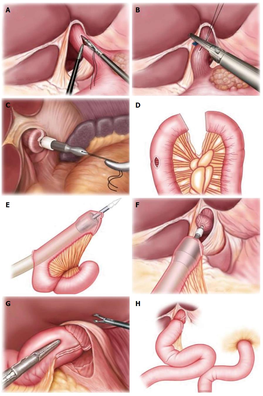
Figure 1 Diagram of gastrointestinal reconstruction after laparoscopic total gastrectomy.
A: The anvil is inserted into the esophagus; B: The esophagus is dissected; C: The anvil is pulled out from the esophagus; D: The jejunum is transected, and a 3-cm longitudinal incision is made at 10-15 cm from the jejunum distal end; E: The circular stapler is inserted into the jejunum; F: Esophagojejunal anastomosis is performed; G: Semi-end-to-end esophagojejunal anastomosis is completed; H: Roux-en-Y anastomosis is completed.
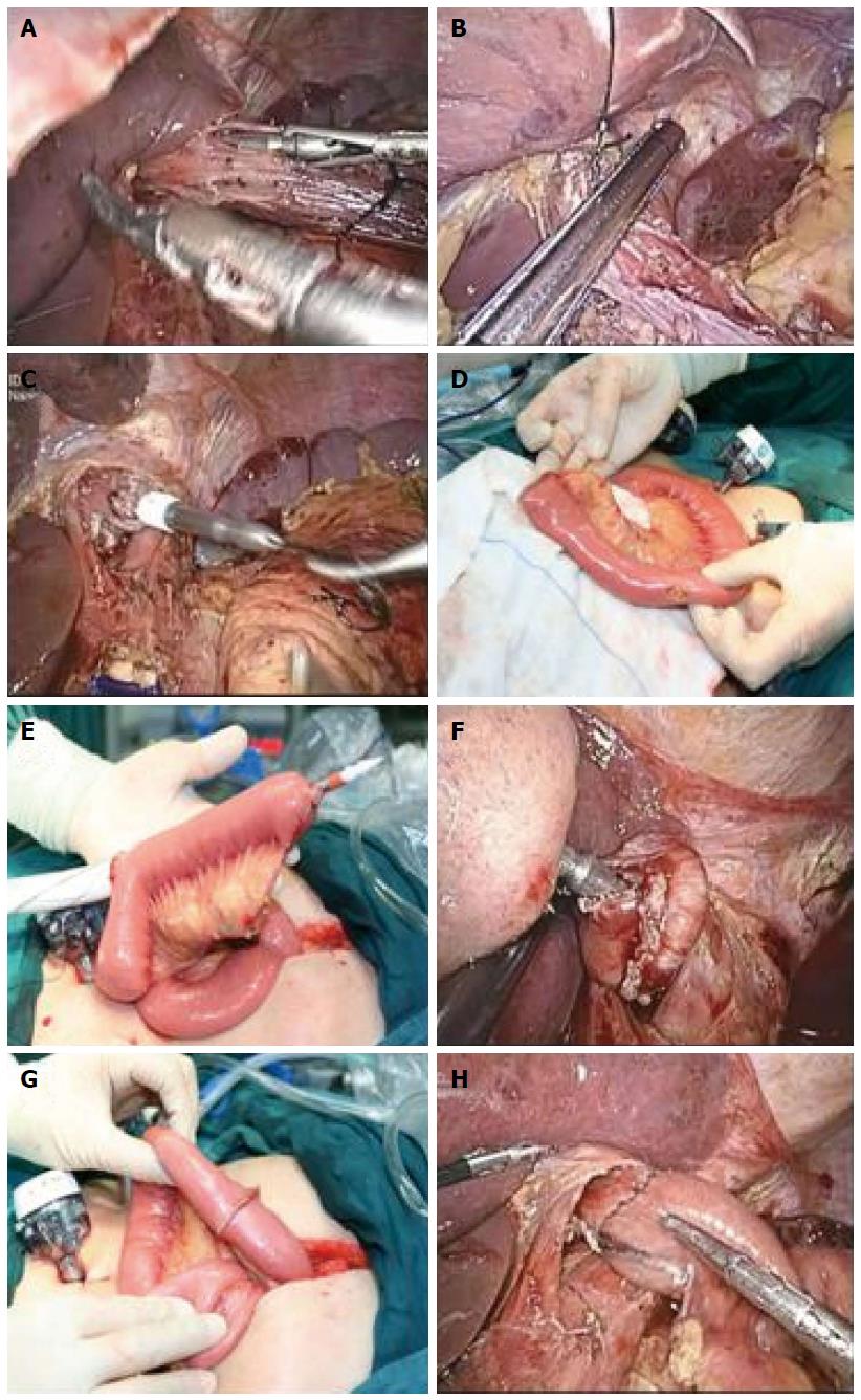
Figure 2 Surgical diagram of esophagojejunal anastomosis after laparoscopic total gastrectomy.
A: The anvil is inserted into the esophagus; B: The esophagus is dissected; C: The anvil is pulled out from the esophagus; D: The jejunum is transected, and a 3-cm longitudinal incision is made at 10-15 cm from the jejunum distal end; E: The circular stapler is inserted into the distal jejunum; F: Esophagojejunal anastomosis is performed; G: Semi-end-to-end esophagojejunal anastomosis is completed; H: The jejunum incision is closed transversely.
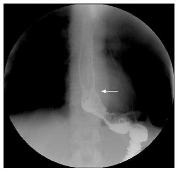
Figure 3 Postoperative Barium meal examination demonstrates normal perfusion after semi-end-to-end esophagojejunal anastomosis.
An open arrow indicates the anastomosis site.
- Citation: Zhao YL, Su CY, Li TF, Qian F, Luo HX, Yu PW. Novel method for esophagojejunal anastomosis after laparoscopic total gastrectomy: Semi-end-to-end anastomosis. World J Gastroenterol 2014; 20(37): 13556-13562
- URL: https://www.wjgnet.com/1007-9327/full/v20/i37/13556.htm
- DOI: https://dx.doi.org/10.3748/wjg.v20.i37.13556


