Copyright
©2014 Baishideng Publishing Group Inc.
World J Gastroenterol. Oct 7, 2014; 20(37): 13382-13401
Published online Oct 7, 2014. doi: 10.3748/wjg.v20.i37.13382
Published online Oct 7, 2014. doi: 10.3748/wjg.v20.i37.13382
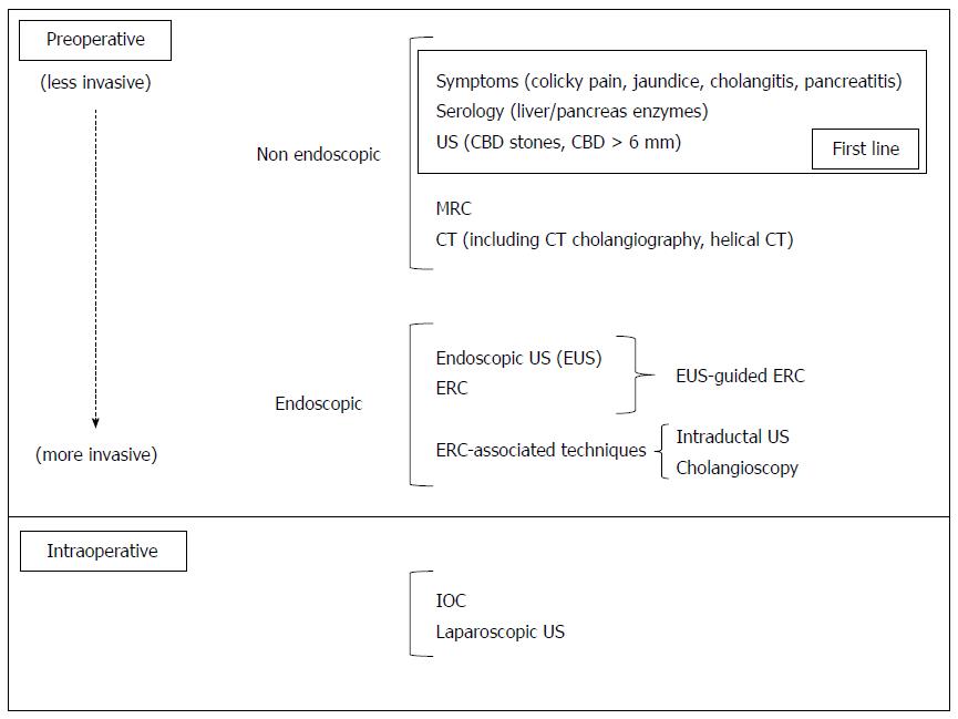
Figure 1 Clinical symptoms, serology and imaging options for the diagnosis of common bile duct stones.
US: Ultrasounds; MRC: Magnetic resonance cholangiography; CT: Computerized tomography; ERC: Endoscopic retrograde cholangiography; IOC: Intraoperative cholangiography; CBD: Common bile duct.
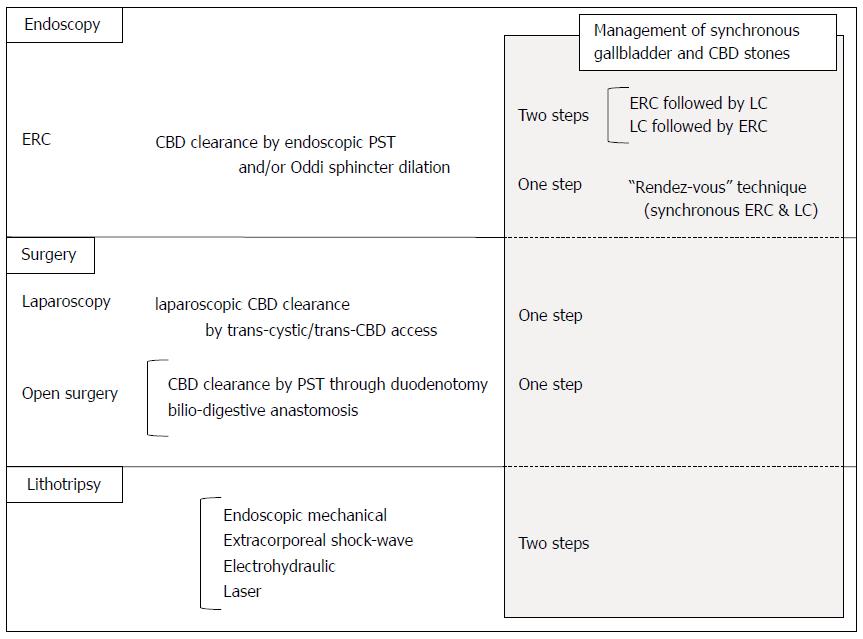
Figure 2 Management options for common bile duct stones.
CBD: Common bile duct; ERC: Endoscopic retrograde cholangiography; LC: Laparoscopic cholecystectomy; PST: Papillosphincterotomy.
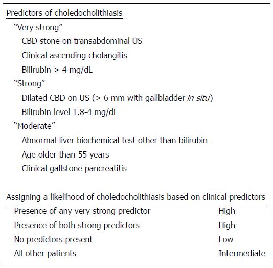
Figure 3 The American Society for Gastrointestinal Endoscopy estimation of risk of carrying common bile duct stones in patients with symptomatic cholelithiasis based on clinical predictors[39].
US: Ultrasounds; CBD: Common bile duct. With the permission of the journal Gastrointestinal Endoscopy.
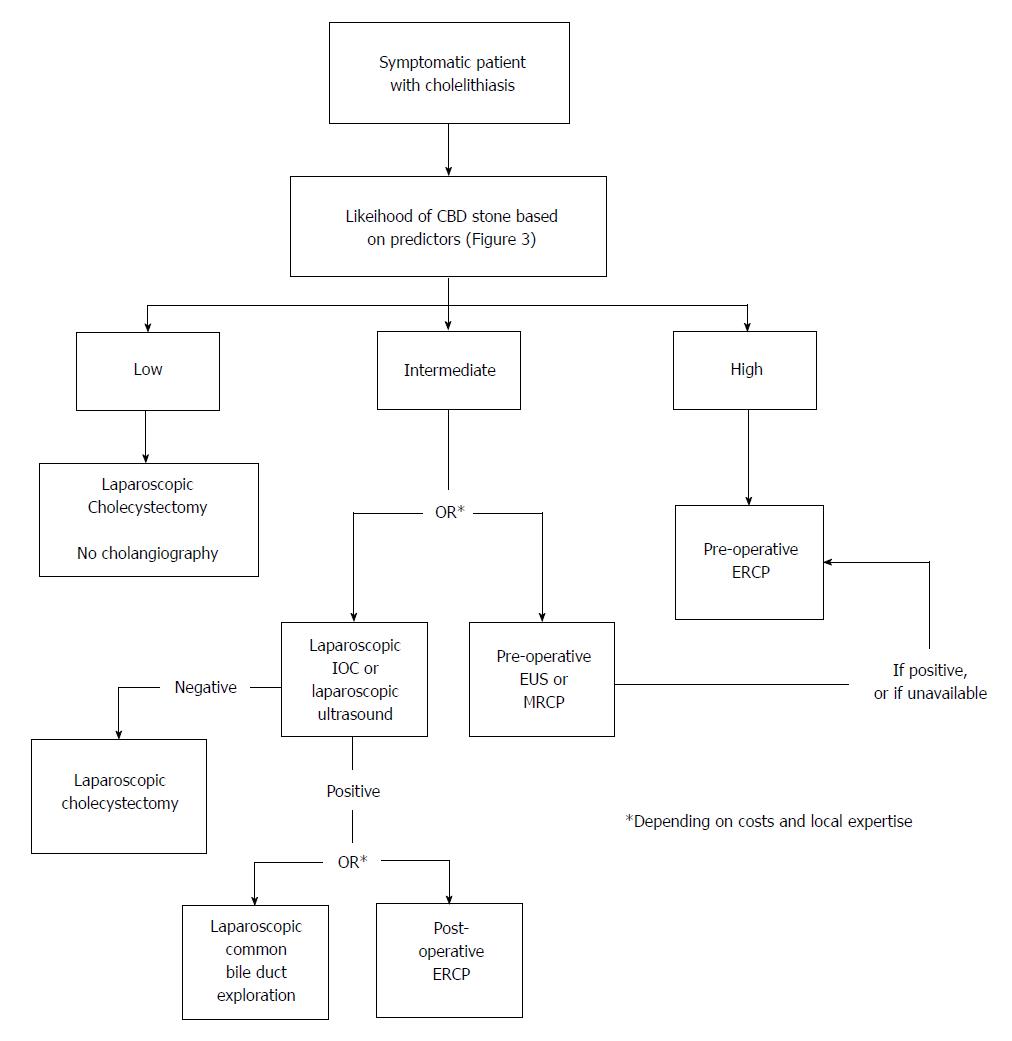
Figure 4 The American Society for Gastrointestinal Endoscopy algorithm for the management of patients with symptomatic cholelithiasis based on the degree of probability for choledocholithiasis[39].
CBD: Common bile duct; IOC: Intraoperative cholangiography; EUS: Endoscopic ultrasounds; MRCP: Magnetic resonance cholangiopancreatographygraphy; ERCP: Endoscopic retrograde cholangiopancreatography. With the permission of the journal Gastrointestinal Endoscopy.
- Citation: Costi R, Gnocchi A, Di Mario F, Sarli L. Diagnosis and management of choledocholithiasis in the golden age of imaging, endoscopy and laparoscopy. World J Gastroenterol 2014; 20(37): 13382-13401
- URL: https://www.wjgnet.com/1007-9327/full/v20/i37/13382.htm
- DOI: https://dx.doi.org/10.3748/wjg.v20.i37.13382


