Copyright
©2014 Baishideng Publishing Group Inc.
World J Gastroenterol. Sep 28, 2014; 20(36): 13139-13145
Published online Sep 28, 2014. doi: 10.3748/wjg.v20.i36.13139
Published online Sep 28, 2014. doi: 10.3748/wjg.v20.i36.13139
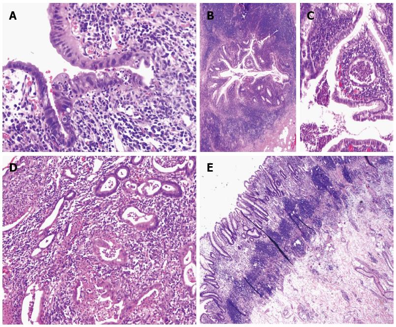
Figure 1 Microscopic features of "ulcerative colitis-like" Crohn’s disease.
A: Acute ileitis with architectural distortion. Section shows prominent villous neutrophilc infiltrate and villous blunting; B and C: A cross-section of appendix shows active inflammation with crypt destruction and crypt abscess. This case only has distal colon involvement. The active appendicitis involves the entire appendix; D: A representative section of the colon shows prominent lamina propria neutrophilic infiltrate associated with ruptured crypt abscess; E: Prominent lymphoid aggregates are present in the superficial and deep submucosa.
- Citation: James SD, Wise PE, Zuluaga-Toro T, Schwartz DA, Washington MK, Shi C. Identification of pathologic features associated with “ulcerative colitis-like” Crohn’s disease. World J Gastroenterol 2014; 20(36): 13139-13145
- URL: https://www.wjgnet.com/1007-9327/full/v20/i36/13139.htm
- DOI: https://dx.doi.org/10.3748/wjg.v20.i36.13139


