Copyright
©2014 Baishideng Publishing Group Inc.
World J Gastroenterol. Sep 28, 2014; 20(36): 12956-12980
Published online Sep 28, 2014. doi: 10.3748/wjg.v20.i36.12956
Published online Sep 28, 2014. doi: 10.3748/wjg.v20.i36.12956
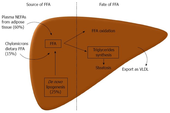
Figure 1 Pathogenesis of liver steatosis.
Hepatic steatosis can result from an increased influx of lipids, free fatty acids (FFA), to the liver or a decreased lipid disposal. Three main sources of FFA in the liver are the plasmatic nonesterified fatty acids (NEFAs), which originate predominantly from lipolysis in the adipose tissue, from de novo lipogenesis, mainly from glucose or other carbohydrates, and from FFA that come in chylomicrons from the gut (dietary FFA). In the liver, FFA can either be oxidized, mainly in the mitochondria, beta-oxidation, or can be used to produce triglycerides. The latter can be exported as very low density lipoproteins (VLDL) to the circulation or can accumulate in lipid droplets in the hepatocyte leading to steatosis.
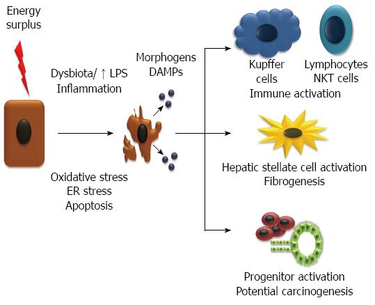
Figure 2 Injury and inflammation leads to non-alcoholic steatohepatitis and fibrogenesis.
Energy surplus leads to fat accumulation in the hepatocytes which promote oxidative stress, endoplasmic reticulum (ER) stress and apoptosis. The injury of hepatocytes is promoted by an inflammatory state, among other factors, favored by a deregulated gut microbiota and increase in lipopolysaccharide (LPS). Injured and dying hepatocytes release damage associated molecular patterns (DAMPs) and morphogens (e.g. hedgehog and Wnt), that act on the immune system increasing inflammation, in stellate cells and progenitors cells activating them and inducing fibrogenesis and pathways of hepatocarcinogenesis. FFA: Free fatty acids; NEFA: Nonesterified fatty acids.
- Citation: Machado MV, Cortez-Pinto H. Non-alcoholic fatty liver disease: What the clinician needs to know. World J Gastroenterol 2014; 20(36): 12956-12980
- URL: https://www.wjgnet.com/1007-9327/full/v20/i36/12956.htm
- DOI: https://dx.doi.org/10.3748/wjg.v20.i36.12956


