Copyright
©2014 Baishideng Publishing Group Inc.
World J Gastroenterol. Aug 28, 2014; 20(32): 11347-11355
Published online Aug 28, 2014. doi: 10.3748/wjg.v20.i32.11347
Published online Aug 28, 2014. doi: 10.3748/wjg.v20.i32.11347
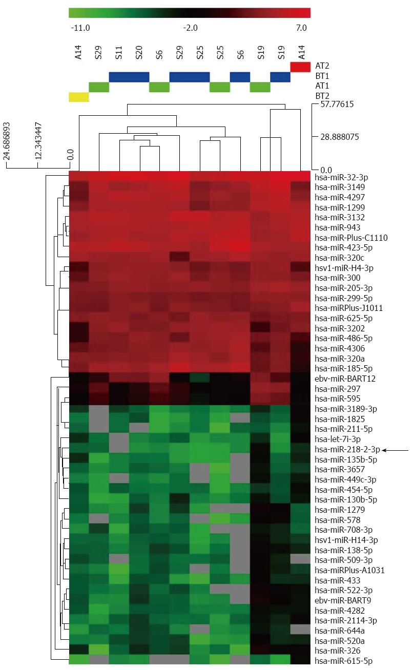
Figure 1 Hierarchical clustering of microRNA in advanced gastric cancer serum samples.
Advanced gastric cancer (AGC) serum samples were clustered according to the expression profile of 86 differentially-expressed microRNAs (miRNAs) of the paired serum samples of 5 AGC patients collected 1 h before and 1 h after cytoreductive surgery plus hyperthermic intraperitoneal chemotherapy. Samples were in columns and miRNAs in rows. The P values for these miRNAs were less than 0.05 in AGC serum samples.
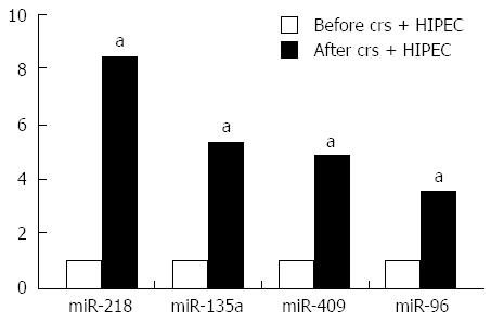
Figure 2 Upregulation of microRNA after cytoreductive surgery and hyperthermic intraperitoneal chemotherapy treatment.
aP < 0.05 vs control.
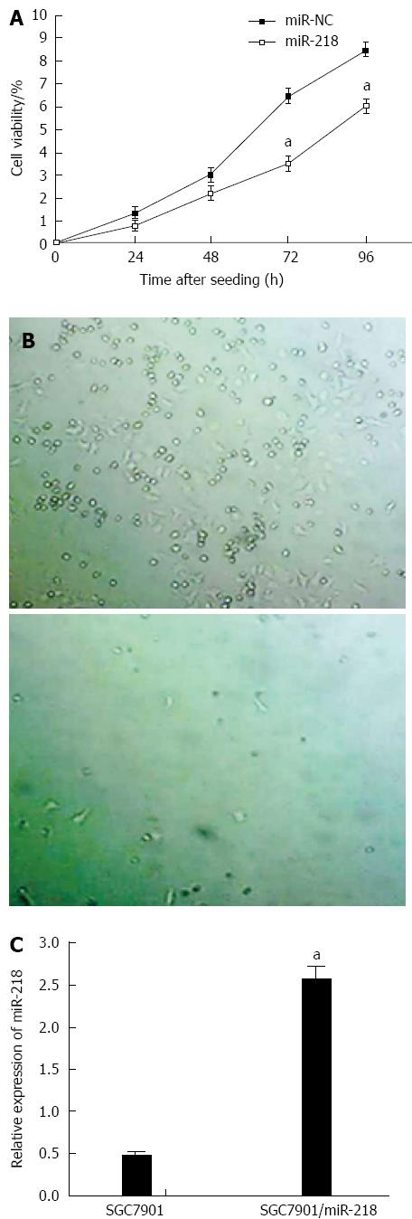
Figure 3 Upregulation of miR-218 inhibited growth of gastric cancers.
A: The upregulation of miR-218 inhibited SGC7901 (human gastric cancer cells) cell growth. Cell viability was measured using a WST-1 kit at indicated time points. Data are presented as mean ± SD from three independent experiments performed in sextuple; B: miR-218 overexpression reduced colony formation of SGC7901 cells. 103 cells were mixed with agarose and seeded in 9-well plates for two weeks; C: The expression levels of miR-218 in the parental SGC7901 and the SGC7901/miR-218 stable cell line. Data are presented as mean ± SD. aP < 0.05 vs control.
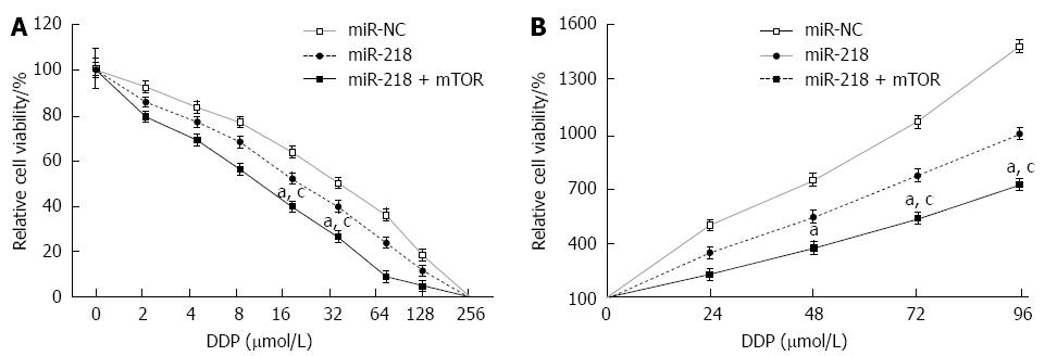
Figure 4 miR-218 increased chemosensitivity to cisplatin could be accelerated by overexpression of mTOR.
Cells were transfected with miR-NC, or miR-218 with or without mTOR cDNA. Cells were exposed to cisplatin for further detection 10 h after transfection. A: Tumor cells proliferation assay of different cisplatin concentrations. 6 × 10³ cells were seeded in 96-well plates and incubated with different concentration of cisplatin for 72 h. Data are presented as mean ± SD from three independent experiments performed in sextuple; B: Cell proliferation in the presence of 12 μmol/L cisplatin. Data are presented as mean ± SD from three independent experiments performed in sextuple. aP < 0.05 vs control; cP < 0.05 vs miR-218 + mTOR.
Figure 5 Upregulation of miR-218 inhibited gastric tumor growth in vivo.
Cells stably expressing miR-218 or miR-NC were incubated in Dulbecco's modified Eagle’s medium and subcutaneously injected into each side of the posterior flank of nude mice (n = 24). Thirty-three days after injection, mice were sacrificed and tumors were removed. A: Tumor volume at 33 d; B: Tumor volumes were detected every three days from the time they were obvious; C: Average tumor weights. aDenoted statistical significance between the two groups of miR-218 and control. aP < 0.05 vs control.
- Citation: Zhang XL, Shi HJ, Wang JP, Tang HS, Wu YB, Fang ZY, Cui SZ, Wang LT. MicroRNA-218 is upregulated in gastric cancer after cytoreductive surgery and hyperthermic intraperitoneal chemotherapy and increases chemosensitivity to cisplatin. World J Gastroenterol 2014; 20(32): 11347-11355
- URL: https://www.wjgnet.com/1007-9327/full/v20/i32/11347.htm
- DOI: https://dx.doi.org/10.3748/wjg.v20.i32.11347


