Copyright
©2014 Baishideng Publishing Group Inc.
World J Gastroenterol. Aug 14, 2014; 20(30): 10637-10641
Published online Aug 14, 2014. doi: 10.3748/wjg.v20.i30.10637
Published online Aug 14, 2014. doi: 10.3748/wjg.v20.i30.10637
Figure 1 Computed tomography imaging.
A: Computed tomography plain scanning showing multiple hyperintense lesions in the spleen; B: During the arterial phase, the lesions showed heterogeneous enhancement; C: In the portal phase, the lesions were more hyperdense than the splenic parenchyma.
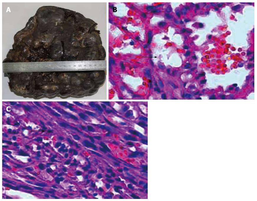
Figure 2 Resected specimen was 2130 g in weight and 24 cm × 19 cm × 11 cm in size.
A: Resected specimen showed splenomegaly with multiple nodular lesions of various sizes; B: Histological section showing disappearance of normal splenic structure, and replacement by various sizes of nodular lesions, which were composed mainly of variably dilated, unorganized vascular channels (black arrow) (original magnification × 200); C: Proliferative spindle cells arranged in fascicular shape (black arrow) (original magnification × 200).
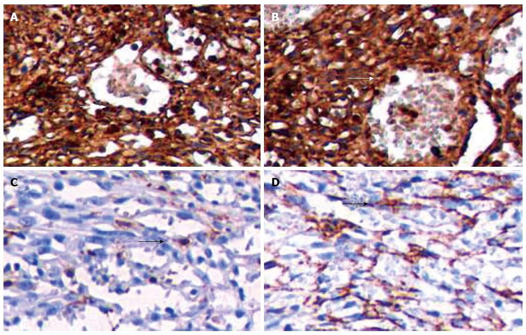
Figure 3 Immunohistochemical staining.
A: CD34 immunostaining was positive in the lining cells (white arrow) and some spindle cells (white arrow) (original magnification × 100); B: Vimentin immunostaining was positive in the tumor (white arrow) (original magnification × 100); C: Factor-VIII-related antigen immunostaining was multifocally positive in the lining cells (black arrow) (original magnification × 100); D: Smooth muscle actin was positive in some spindle cells (black arrow) (original magnification × 100).
- Citation: Wang RT, Xu XS, Hou HL, Qu K, Bai JG. Symptomatic multinodular splenic hamartoma preoperatively suspected as metastatic tumor: A case report. World J Gastroenterol 2014; 20(30): 10637-10641
- URL: https://www.wjgnet.com/1007-9327/full/v20/i30/10637.htm
- DOI: https://dx.doi.org/10.3748/wjg.v20.i30.10637


