Copyright
©2014 Baishideng Publishing Group Inc.
World J Gastroenterol. Aug 14, 2014; 20(30): 10553-10563
Published online Aug 14, 2014. doi: 10.3748/wjg.v20.i30.10553
Published online Aug 14, 2014. doi: 10.3748/wjg.v20.i30.10553
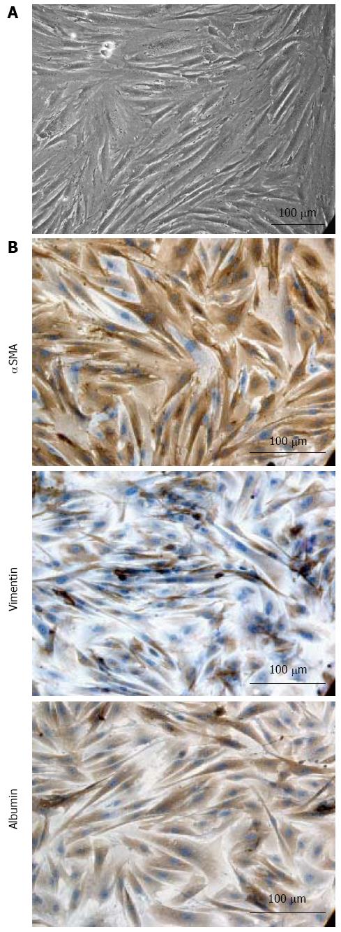
Figure 1 Adult-derived human liver stem/progenitor cells characterization.
A: Morphology of adult-derived human liver stem/progenitor cells (ADHLSC) in expansion medium. Phase contrast microscopic view of ADHLSC obtained after second passage. Magnification: × 100; B: Immunocytochemistry characterization (passage 6) analysis: expression of mesenchymal and hepatic markers in ADHLSC. Mesenchymal markers: α-smooth muscle actin (αSMA) and vimentin and hepatic marker albumin immunostaining were analyzed by direct light microscopy. ADHLSC plated on collagen type I-coated coverslips were fixed and incubated with corresponding primary antibodies. Immunoreactivity was visualized using horseradish peroxydase (HRP) Cell nuclei are counterstaining with Mayer’s hematoxylin (blue). Images are representative of several fields examined.
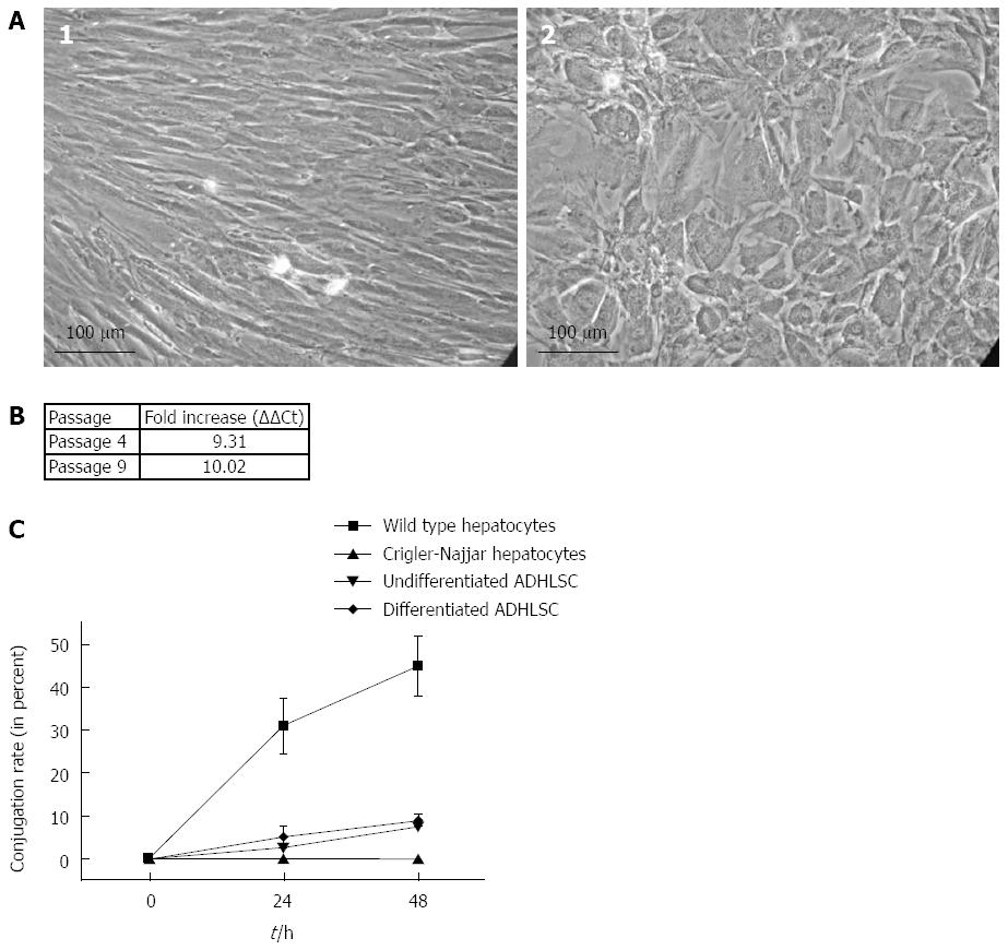
Figure 2 Hepatogenic differentiation and expression of functional uridine diphosphate-glucuronosyltransferase 1A1.
A: Evaluation of in vitro adult-derived human liver stem/progenitor cells (ADHLSC) hepatic differentiation potential at passage 6. Undifferentiated ADHLSC (1) were able to change their morphology by adopting a polygonal shape with a granular cytoplasm after 4 wk of treatment with specific growth factors and cytokines. Magnification: × 100; B: Uridine diphosphate-glucuronosyltransferase 1A (UGT1A) mRNA relative expression quantification. Results were obtained after passage 4 and 9. UGT1A mRNA level in differentiated ADHLSC, were normalized to Cyclophilin A and reported to undifferentiated ADHLSC UGT1A mRNA level; C: In vitro bilirubin conjugation kinetic assessment; Cells were incubated with 100 μmol/L unconjugated bilirubin, supernatants were harvested after 24 h or 48 h. Conjugation rate (CR) was evaluated with high-performance liquid chromatography as follow: [(Conjugated bilirubin concentration)/(Total bilirubin concentration)] x 100. Squares: Wild type hepatocytes (positive control); Triangle: Crigler-Najjar patient hepatocytes (negative control), overturned triangle: undifferentiated ADHLSC, rhomb: differentiated ADHLSC.
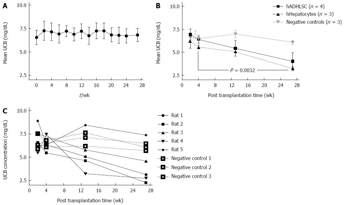
Figure 3 Injected Gunn rat follow up.
A: Assessment of bilirubin concentration stability in 11 naive rats of the Gunn rat colony followed during 27 wk. Bilirubin concentration is expressed in mg/dL; B: Mean of unconjugated bilirubin for adult-derived human liver stem/progenitor cells (ADHLSC) injected rats n = 4 (black line), human hepatocytes injected rats n = 3 and for negative control n = 3 (grey line). Difference between 2 wk and 27 wk post injection concentration bilirubin was significant for ADHLSC injected rats (P = 0.0032). Mean of unconjugated bilirubin (UCB) concentration is expressed in mg/dL. Squares: hADHLSC; Triangles: human hepatocytes; Inverted triangles: negative controls; C: Unconjugated bilirubin evolution with time. Black lines: ADHLSC transplanted Gunn rats n = 5; dotted lines: PBS injected rats n = 3 (negative control). Bilirubin concentration is expressed in mg/dL.
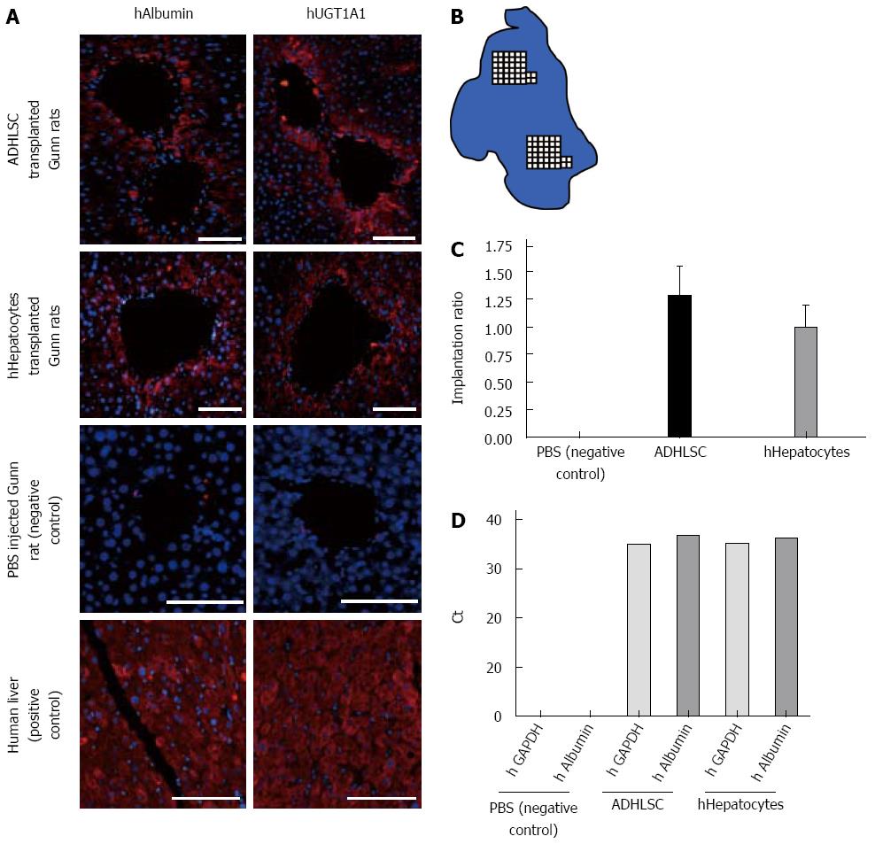
Figure 4 Liver analysis and human cells quantification.
A: Immunohistochemistry against human albumin and human uridine diphosphate-glucuronosyltransferase 1A1 (UGT1A1) proteins on serial liver slices in Gunn rats transplanted with adult-derived human liver stem/progenitor cells (ADHLSC) and hHepatocytes. Immunostainings were analyzed by fluorescent microscopy. Slices were fixed and incubated with corresponding primary antibodies. Immunoreactivity was visualized using a 1/500 AlexaFluor594 anti-rabbit antibody solution. Human liver was used as positive control for albumin and UGT1A1 staining whereas negative control consisted of Gunn rat liver injected with PBS. Cell nuclei are stained using DAPI (blue). Bar scale: 200 μm; B: Schematic representation of an immunostained slide. Human albumin positive cells were counted over two randomly chosen areas of the section; C: Mean of human cells repopulation ratio in PBS (negative control), ADHLSC (black column) and hHepatocytes (grey column) transplanted Gunn rats (P > 0.05). Measurement was based on human albumin positive cells counted over two randomly chosen areas of the section; D: Human GAPDH (light grey column) and albumin (dark grey column) detection by RTqPCR in negative control (PBS injected), ADHLSC, and human hepatocyte injected rat livers.
- Citation: Maerckx C, Tondreau T, Berardis S, Pelt JV, Najimi M, Sokal E. Human liver stem/progenitor cells decrease serum bilirubin in hyperbilirubinemic Gunn rat. World J Gastroenterol 2014; 20(30): 10553-10563
- URL: https://www.wjgnet.com/1007-9327/full/v20/i30/10553.htm
- DOI: https://dx.doi.org/10.3748/wjg.v20.i30.10553


