Copyright
©2014 Baishideng Publishing Group Inc.
World J Gastroenterol. Jul 28, 2014; 20(28): 9314-9320
Published online Jul 28, 2014. doi: 10.3748/wjg.v20.i28.9314
Published online Jul 28, 2014. doi: 10.3748/wjg.v20.i28.9314
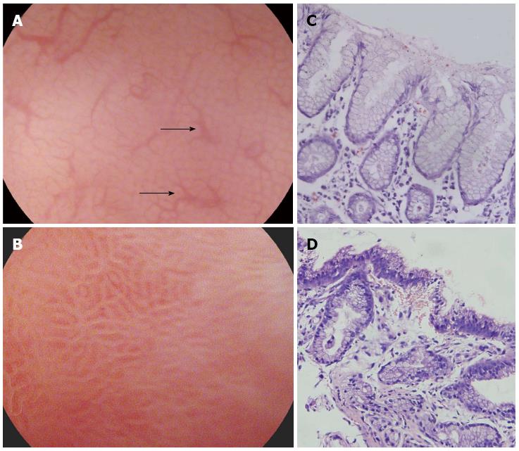
Figure 1 Gastric body mucosal patterns on magnifying endoscopy.
A: Normal mucosa comprises a honeycomb−type subepithelial capillary network (SECN) with a regular arrangement of collecting venules (arrow) and regular, round pits; B: Helicobacter pylori (H. pylori)-associated gastritis appears loss of the normal SECN and collecting venules, with enlarged white pits surrounded by erythema; C: Corresponding histologic features of Figure 1A; D: Corresponding histologic features of Figure 1B.
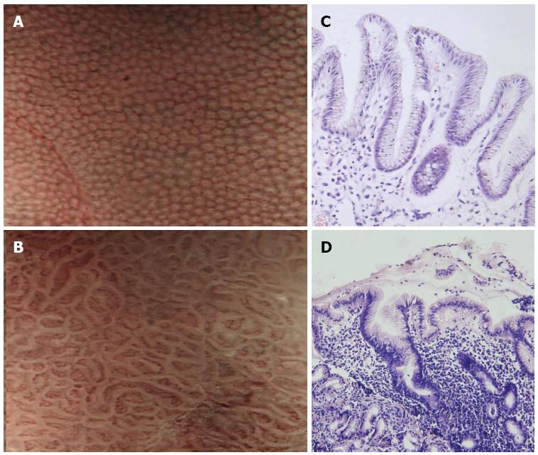
Figure 2 Narrow band imaging of the gastric body.
A: Normal mucosa shows uniform small, round pits surrounded by honeycomb-like subepithelial capillary networks (SECNs); B: Helicobacter pylori (H. pylori)-associated gastritis shows obviously enlarged, oval or prolonged pits; C: Corresponding histologic features of Figure 2A; D: Corresponding histologic features of Figure 2B.
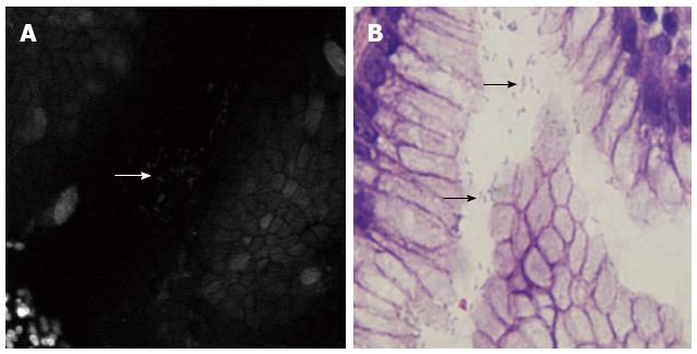
Figure 3 Comparison of confocal laser endomicroscopy images and histology of surface gastric mucosa.
A: Accumulated white spots (arrow) visible on the epithelial layer and gastric pits after topical application of acriflavine; B: Histopathology specimens (arrows).
-
Citation: Ji R, Li YQ. Diagnosing
Helicobacter pylori infectionin vivo by novel endoscopic techniques. World J Gastroenterol 2014; 20(28): 9314-9320 - URL: https://www.wjgnet.com/1007-9327/full/v20/i28/9314.htm
- DOI: https://dx.doi.org/10.3748/wjg.v20.i28.9314


