Copyright
©2014 Baishideng Publishing Group Inc.
World J Gastroenterol. Jul 21, 2014; 20(27): 9205-9209
Published online Jul 21, 2014. doi: 10.3748/wjg.v20.i27.9205
Published online Jul 21, 2014. doi: 10.3748/wjg.v20.i27.9205
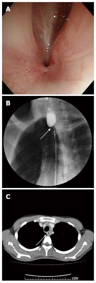
Figure 1 Preoperative findings.
A: An upper gastrointestinal endoscopy revealed a pin-hole like stricture that was located 19 cm distally from the incisors; B: An esophagography revealed circumferential stenosis of the esophagus extending distally 19 cm from the incisors to the esophagogastric junction (arrow); C: A Computed tomography detected circumferential stenosis and wall thickness at the same site (arrow).
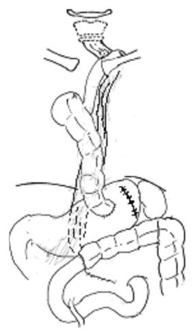
Figure 2 Design of the surgical reconstruction performed via the retrosternal route using ileocolon interposition.
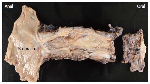
Figure 3 Gross findings of the resected specimen.
Remarkable wall thickness was observed throughout the entire length of the esophagus, and the luminal area was distinctively stenosed, particularly at the esophagogastric junction. The oral side of the specimen was additionally resected because of the pin-hole like stricture detected at the proximal margin.
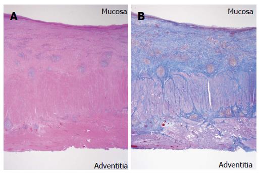
Figure 4 Histopathological findings.
A: Massive fibrosis and infiltration of plasmacytes and lymphocytes were primarily detected in the submucosa throughout the esophagus (hematoxylin and eosin stain (H and E); original magnification, × 11); B: Massive fibrosis extended to the muscularis propria and adventitia along nearly all of the esophagus. The lumen was largely covered by regenerative squamous epithelium with scattered erosion (Masson`s trichrome stain; original magnification, × 11).
- Citation: Kitajima T, Momose K, Lee S, Haruta S, Shinohara H, Ueno M, Fujii T, Udagawa H. Benign esophageal stricture after thermal injury treated with esophagectomy and ileocolon interposition. World J Gastroenterol 2014; 20(27): 9205-9209
- URL: https://www.wjgnet.com/1007-9327/full/v20/i27/9205.htm
- DOI: https://dx.doi.org/10.3748/wjg.v20.i27.9205


