Copyright
©2014 Baishideng Publishing Group Inc.
World J Gastroenterol. Jul 21, 2014; 20(27): 9205-9209
Published online Jul 21, 2014. doi: 10.3748/wjg.v20.i27.9205
Published online Jul 21, 2014. doi: 10.3748/wjg.v20.i27.9205
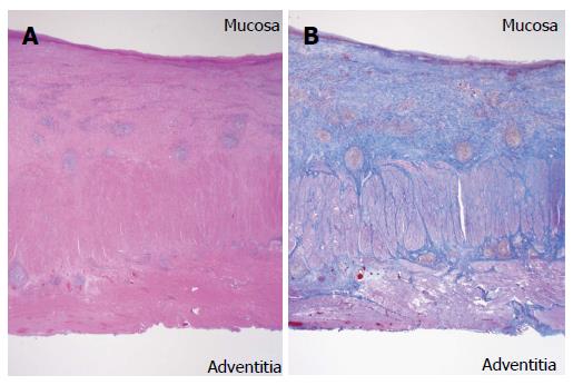
Figure 4 Histopathological findings.
A: Massive fibrosis and infiltration of plasmacytes and lymphocytes were primarily detected in the submucosa throughout the esophagus (hematoxylin and eosin stain (H and E); original magnification, × 11); B: Massive fibrosis extended to the muscularis propria and adventitia along nearly all of the esophagus. The lumen was largely covered by regenerative squamous epithelium with scattered erosion (Masson`s trichrome stain; original magnification, × 11).
- Citation: Kitajima T, Momose K, Lee S, Haruta S, Shinohara H, Ueno M, Fujii T, Udagawa H. Benign esophageal stricture after thermal injury treated with esophagectomy and ileocolon interposition. World J Gastroenterol 2014; 20(27): 9205-9209
- URL: https://www.wjgnet.com/1007-9327/full/v20/i27/9205.htm
- DOI: https://dx.doi.org/10.3748/wjg.v20.i27.9205


