Copyright
©2014 Baishideng Publishing Group Inc.
World J Gastroenterol. Jul 7, 2014; 20(25): 8312-8316
Published online Jul 7, 2014. doi: 10.3748/wjg.v20.i25.8312
Published online Jul 7, 2014. doi: 10.3748/wjg.v20.i25.8312
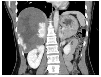
Figure 1 There was contrast enhancement at the periphery of the mass consistent with a cavernous hemangioma.
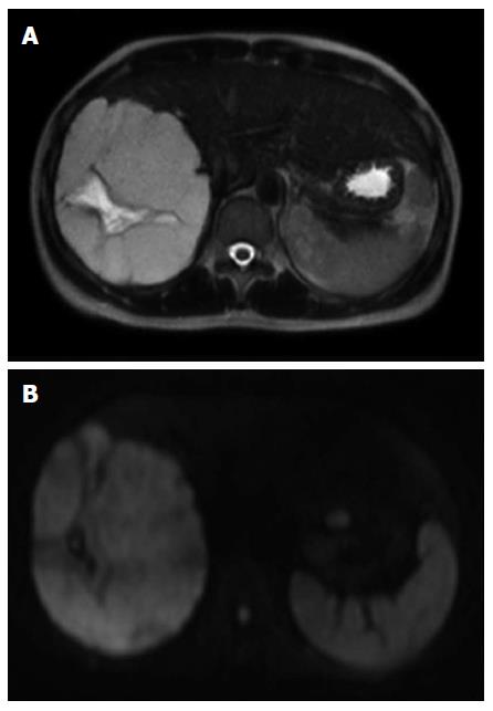
Figure 2 High intensity on T2-weighted magnetic resonance imaging (A) and high intensity on diffusion magnetic resonance imaging (B).
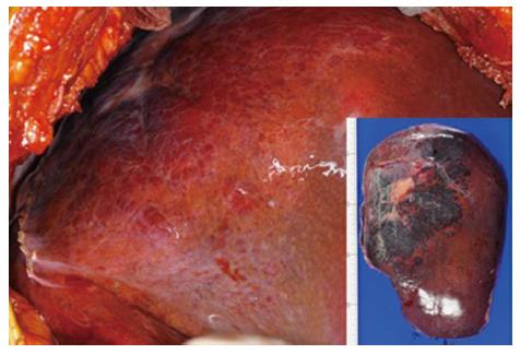
Figure 3 Resected tumor was 200 mm × 140 mm × 85 mm in size and 910 g in weight.
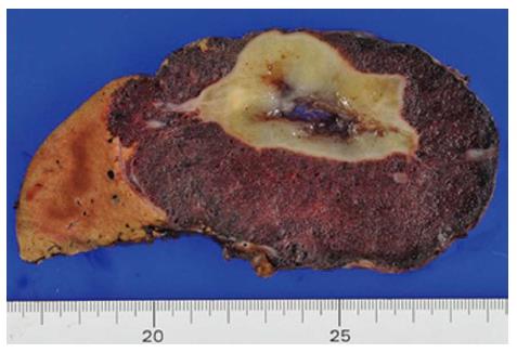
Figure 4 Cut section of the resected tumor.
The white area has tumor degeneration and loss of tumor cells.
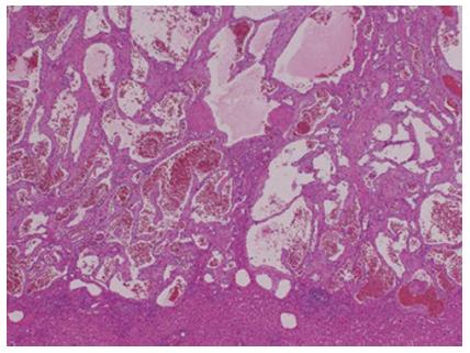
Figure 5 Histological findings of a giant tumor reveal endothelial cell proliferation and dilated blood channels (hematoxylin and eosin staining, × 4).
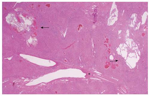
Figure 6 Hemangiomatous lesions (arrows) were scattered around the Glisson’s capsule (star) on the back ground liver (hematoxylin and eosin staining, × 1.
25).
- Citation: Ohkura Y, Hashimoto M, Lee S, Sasaki K, Matsuda M, Watanabe G. Right hepatectomy for giant cavernous hemangioma with diffuse hemangiomatosis around Glisson’s capsule. World J Gastroenterol 2014; 20(25): 8312-8316
- URL: https://www.wjgnet.com/1007-9327/full/v20/i25/8312.htm
- DOI: https://dx.doi.org/10.3748/wjg.v20.i25.8312


