Copyright
©2014 Baishideng Publishing Group Inc.
World J Gastroenterol. Jun 28, 2014; 20(24): 7984-7987
Published online Jun 28, 2014. doi: 10.3748/wjg.v20.i24.7984
Published online Jun 28, 2014. doi: 10.3748/wjg.v20.i24.7984
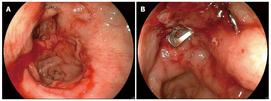
Figure 1 Anastomosis after low anterior resection.
A: Colonoscopy shows breakdown at the anastomotic ring; B: Breakdown site has been closed by the over-the-scope-clipping system.
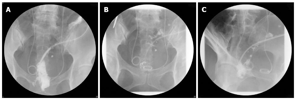
Figure 2 Contrast radiography after low anterior resection.
A: Large abscess is seen outside the bowel; B: Breakdown site has been closed by the over-the-scope-clipping (OTSC) system; C: Abscess has shrunk two days after closure by the OTSC system.
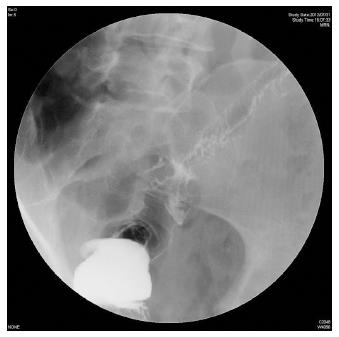
Figure 3 Contrast radiography after sigmoidectomy.
A minor leak is shown.
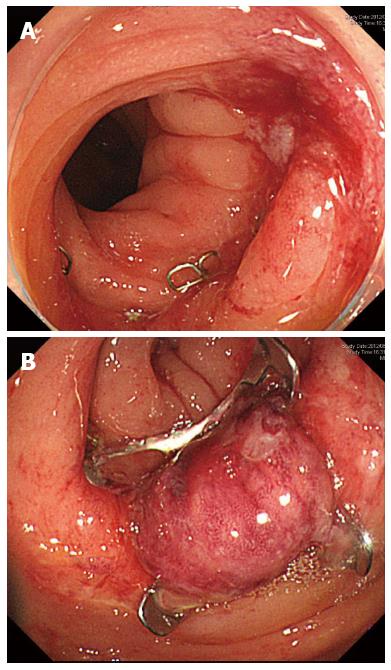
Figure 4 Anastomosis after sigmoidectomy.
A: Colonoscopy shows a reddened area at the anastomosis; B: Reddened area has been closed by the over-the-scope-clipping system.
- Citation: Kobayashi H, Kikuchi A, Okazaki S, Ishiguro M, Ishikawa T, Iida S, Uetake H, Sugihara K. Over-the-scope-clipping system for anastomotic leak after colorectal surgery: Report of two cases. World J Gastroenterol 2014; 20(24): 7984-7987
- URL: https://www.wjgnet.com/1007-9327/full/v20/i24/7984.htm
- DOI: https://dx.doi.org/10.3748/wjg.v20.i24.7984


