Copyright
©2014 Baishideng Publishing Group Inc.
World J Gastroenterol. Jun 14, 2014; 20(22): 7067-7074
Published online Jun 14, 2014. doi: 10.3748/wjg.v20.i22.7067
Published online Jun 14, 2014. doi: 10.3748/wjg.v20.i22.7067
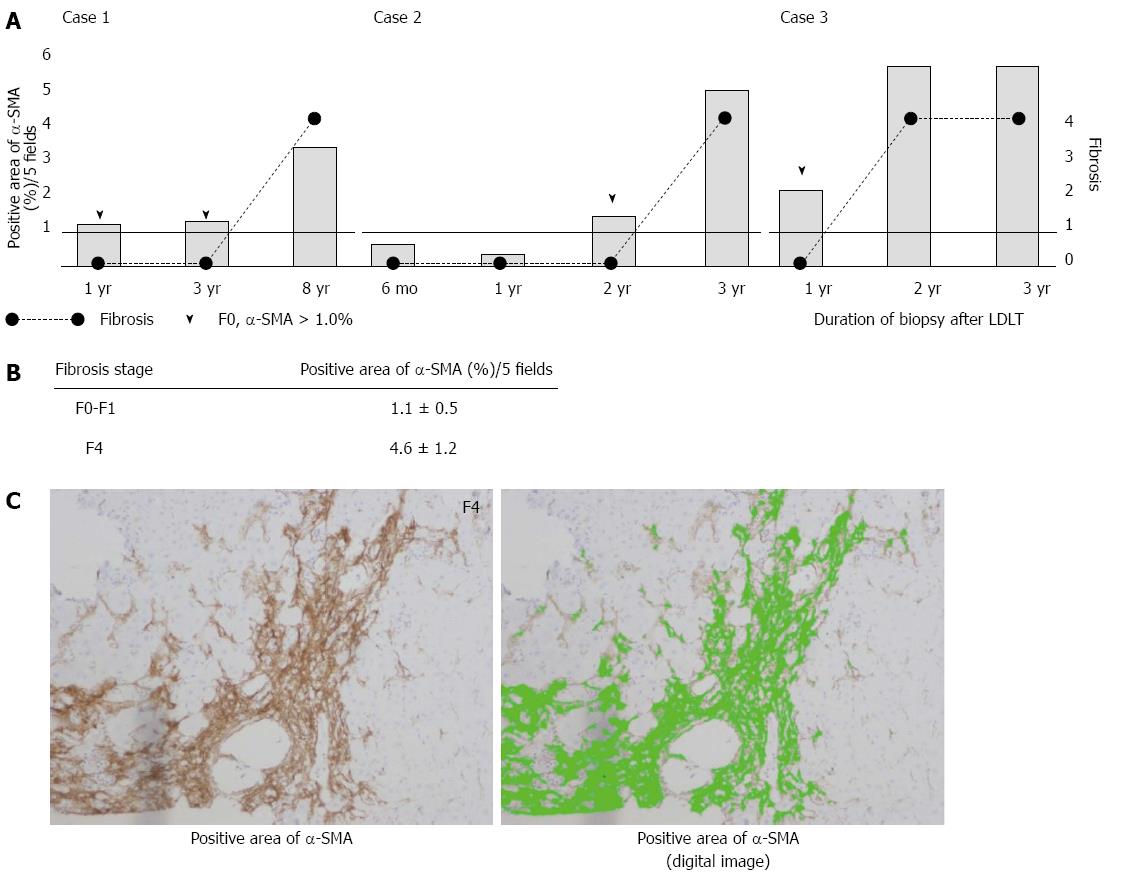
Figure 1 Changes in alpha smooth muscle actin expression in adult patients with fibrosis.
A: The α-smooth muscle actin (SMA)-positive area continued to increase over time in all the patients, even when they were in the pre-fibrotic stage (arrow head); B: The α-SMA positive area ratio in the patients with fibrosis was calculated based on the fibrosis stage. The α-SMA area ratio was higher in the patients with F0-1 fibrosis than in those with F4 fibrosis; C: The photograph on the left shows the α-SMA staining in a representative case with F4 fibrosis. The photograph on the right shows a WinRoof digital image, with green corresponding to the area of α-SMA-positive staining. α-SMA: Alpha smooth muscle actin; LDLT: Living donor liver transplantation.
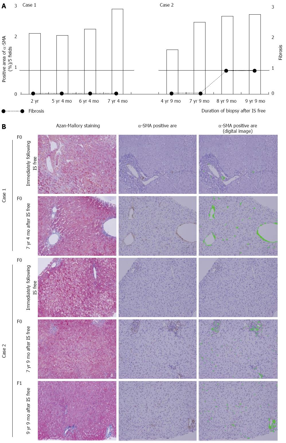
Figure 2 Change in alpha smooth muscle actin expression in the two pediatric cases with immune tolerance.
A: The α-SMA-positive area continued to increase over time in both cases. Case 1 showed F0 fibrosis in the liver at all time points, whereas Case 2 showed a slight progression of fibrosis (F1) eight years after the cessation of immunosuppressive treatment; B: The findings of Azan-Mallory staining and the α-SMA-positive area determined by immunohistochemical analysis are shown. α-SMA: Alpha smooth muscle actin; IS: Immunosuppression.
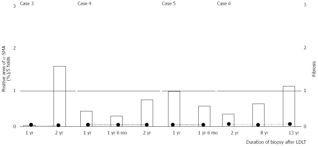
Figure 3 Changes in alpha smooth muscle actin expression in the four pediatric cases that continued immunosuppression.
There was no clear increase in the α-SMA-positive area. α-SMA: Alpha smooth muscle actin; LDLT: Living donor liver transplantation.
- Citation: Hirabaru M, Mochizuki K, Takatsuki M, Soyama A, Kosaka T, Kuroki T, Shimokawa I, Eguchi S. Expression of alpha smooth muscle actin in living donor liver transplant recipients. World J Gastroenterol 2014; 20(22): 7067-7074
- URL: https://www.wjgnet.com/1007-9327/full/v20/i22/7067.htm
- DOI: https://dx.doi.org/10.3748/wjg.v20.i22.7067


