Copyright
©2014 Baishideng Publishing Group Inc.
World J Gastroenterol. Jun 14, 2014; 20(22): 6821-6825
Published online Jun 14, 2014. doi: 10.3748/wjg.v20.i22.6821
Published online Jun 14, 2014. doi: 10.3748/wjg.v20.i22.6821
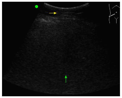
Figure 1 Bedside ultrasound image displaying sonographic characteristics on non-alcoholic fatty liver disease.
Attenuation of image (green arrow), diffuse echogenicity, uniform heterogeneous liver, thick subcutaneous depth (yellow arrow), and enlarged liver filling of the entire field as described by Riley et al[9].
Figure 2 Diagnostic algorithm for suspected non-alcoholic fatty liver disease.
The algorithm illustrates the use of ultrasound in reducing the need for liver biopsy in the diagnosis of non-alcoholic fatty liver disease. BMI: Body mass index; AST/ALT: Aspartate aminotransferase/alanine aminotransferase.
- Citation: Khov N, Sharma A, Riley TR. Bedside ultrasound in the diagnosis of nonalcoholic fatty liver disease. World J Gastroenterol 2014; 20(22): 6821-6825
- URL: https://www.wjgnet.com/1007-9327/full/v20/i22/6821.htm
- DOI: https://dx.doi.org/10.3748/wjg.v20.i22.6821


