Copyright
©2014 Baishideng Publishing Group Inc.
World J Gastroenterol. May 28, 2014; 20(20): 6353-6356
Published online May 28, 2014. doi: 10.3748/wjg.v20.i20.6353
Published online May 28, 2014. doi: 10.3748/wjg.v20.i20.6353
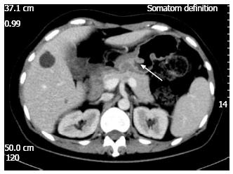
Figure 1 In computed tomography, arrow showed thickened gastric antrum.
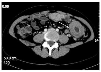
Figure 2 In computed tomography, arrow showed thickened small intestine wall in the left mid-abdomen with peripheral lymph nodes enlargement.
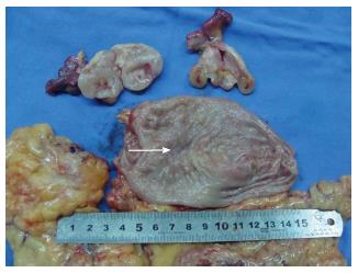
Figure 3 Specimen of stomach and bilateral ovary.
Arrow showed the lesion of gastric antrum.
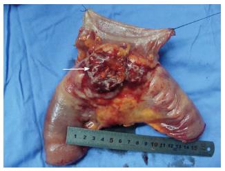
Figure 4 Specimen of segment of small intestine and descending colon.
Arrow showed the lesion of small intestine.
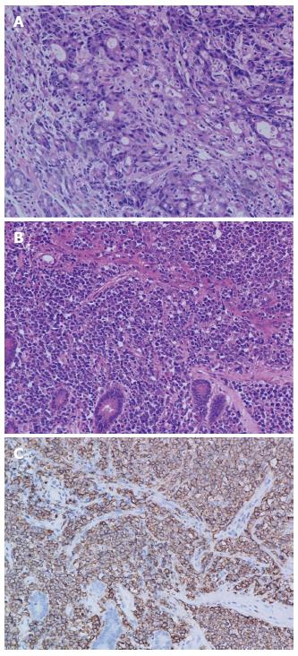
Figure 5 Histopathological finding.
A: Histopathological finding of gastric cancer [hematoxylin and eosin (HE) stain, × 200]; B: Histopathological finding of small intestinal diffuse large B cell lymphoma (HE stain, × 200); C: Immunohistochemical stain was positive with CD20 for small intestinal lymphoma (× 200).
- Citation: Chen DW, Pan Y, Yan JF, Mou YP. Laparoscopic resection of synchronous gastric cancer and primary small intestinal lymphoma: A case report. World J Gastroenterol 2014; 20(20): 6353-6356
- URL: https://www.wjgnet.com/1007-9327/full/v20/i20/6353.htm
- DOI: https://dx.doi.org/10.3748/wjg.v20.i20.6353


