Copyright
©2014 Baishideng Publishing Group Inc.
World J Gastroenterol. May 28, 2014; 20(20): 5987-5998
Published online May 28, 2014. doi: 10.3748/wjg.v20.i20.5987
Published online May 28, 2014. doi: 10.3748/wjg.v20.i20.5987
Figure 1 Radiofrequency ablation combined with right hemihepatectomy for multifocal hepatocellular carcinoma in a 69-year-old woman.
A: Preoperative contrast-enhanced transverse helical computed tomography (CT) scan obtained during the venous phase shows one small hepatocellular carcinoma (HCC) 1.4 cm diameter in the left hepatic lobe (black arrow); B: An HCC 7.0 cm in diameter (black arrow) is present in the right hepatic lobe; C: Contrast-enhanced CT showed round ablation zones (white arrow) 6 mo after resection of the large tumor and concurrent radiofrequency ablation for the small tumor. Tumor recurrence was not found in the remnant liver.
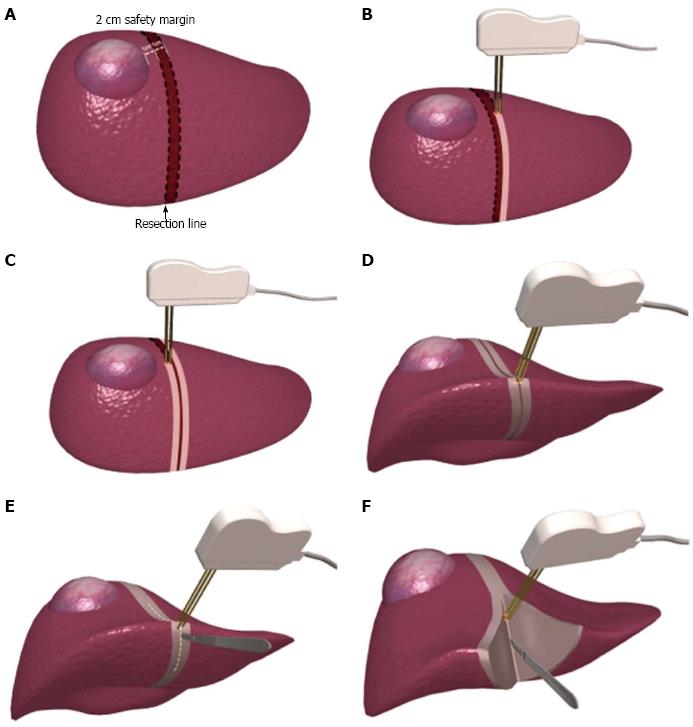
Figure 2 Classic operative technique using a bipolar radiofrequency device for hepatectomy (A-F).
- Citation: Feng K, Ma KS. Value of radiofrequency ablation in the treatment of hepatocellular carcinoma. World J Gastroenterol 2014; 20(20): 5987-5998
- URL: https://www.wjgnet.com/1007-9327/full/v20/i20/5987.htm
- DOI: https://dx.doi.org/10.3748/wjg.v20.i20.5987


