Copyright
©2014 Baishideng Publishing Group Co.
World J Gastroenterol. May 7, 2014; 20(17): 5135-5140
Published online May 7, 2014. doi: 10.3748/wjg.v20.i17.5135
Published online May 7, 2014. doi: 10.3748/wjg.v20.i17.5135
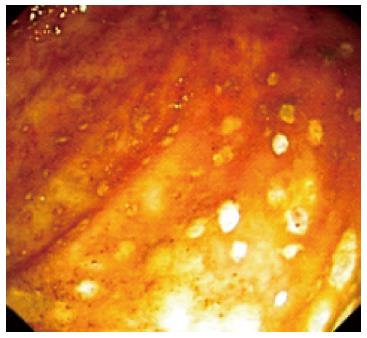
Figure 1 Pseudomembranous colitis in the ulcerative colitis patient.
Colonoscopy image showing pseudomembranes in the descending colon, with a loss mucosal vascular pattern.
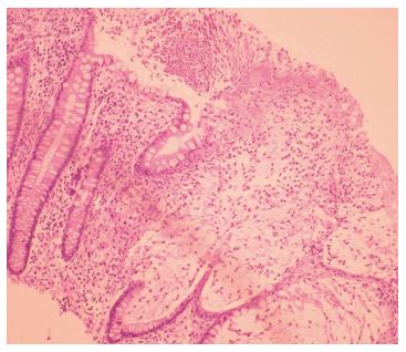
Figure 2 Colonic histology of Clostridium difficile infection superimposed on ulcerative colitis.
Necrosis of superficial crypts with a dense infiltrate of neutrophils, fibrin, and cellular debris covering the mucosal surface.
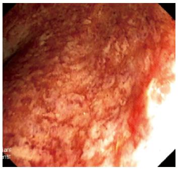
Figure 3 Severe endoscopic aspect of ulcerative colitis.
Ulcerations, loss of vascular pattern and edema of the mucosae were noted in the rectum.
-
Citation: Seicean A, Moldovan-Pop A, Seicean R. Ulcerative colitis worsened after
Clostridium difficile infection: Efficacy of infliximab. World J Gastroenterol 2014; 20(17): 5135-5140 - URL: https://www.wjgnet.com/1007-9327/full/v20/i17/5135.htm
- DOI: https://dx.doi.org/10.3748/wjg.v20.i17.5135


