Copyright
©2014 Baishideng Publishing Group Co.
World J Gastroenterol. Apr 28, 2014; 20(16): 4827-4829
Published online Apr 28, 2014. doi: 10.3748/wjg.v20.i16.4827
Published online Apr 28, 2014. doi: 10.3748/wjg.v20.i16.4827
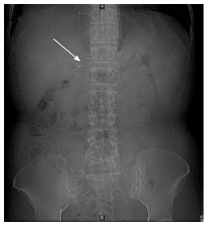
Figure 1 Plain abdominal radiograph showed metal endoclips (arrow) in the right upper quadrant area.
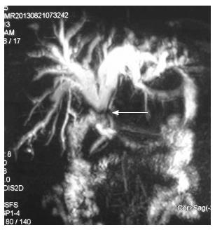
Figure 2 Magnetic resonance cholangiography showed marked dilatation of biliary duct and stenosis of the common bile duct at the hepatic duct confluence (arrow), which was close to the duodenum.
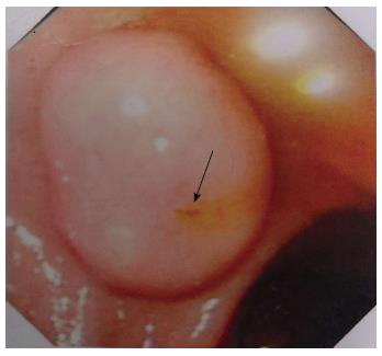
Figure 3 Endoscopic image of the duodenum showed yellowish bile acid (arrow) leaking from a papillary orifice at the first part of duodenum wall.
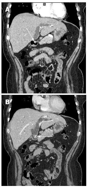
Figure 4 Computed tomography showed a mass on the duodenal wall (arrow), and linear, highly dense lesions both in the mass (A, arrow) and in the hepatic duct confluence (B, arrow) with dilated hepatic ducts.
- Citation: Hong T, Xu XQ, He XD, Qu Q, Li BL, Zheng CJ. Choledochoduodenal fistula caused by migration of endoclip after laparoscopic cholecystectomy. World J Gastroenterol 2014; 20(16): 4827-4829
- URL: https://www.wjgnet.com/1007-9327/full/v20/i16/4827.htm
- DOI: https://dx.doi.org/10.3748/wjg.v20.i16.4827


