Copyright
©2014 Baishideng Publishing Group Co.
World J Gastroenterol. Apr 28, 2014; 20(16): 4822-4826
Published online Apr 28, 2014. doi: 10.3748/wjg.v20.i16.4822
Published online Apr 28, 2014. doi: 10.3748/wjg.v20.i16.4822
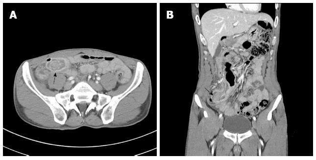
Figure 1 Preoperative abdominal computed tomography findings.
“Target sign”, which can be shown from intussusception, was identified in the terminal ileum (black arrow). A: Horizontal plane; B: Coronal plane.
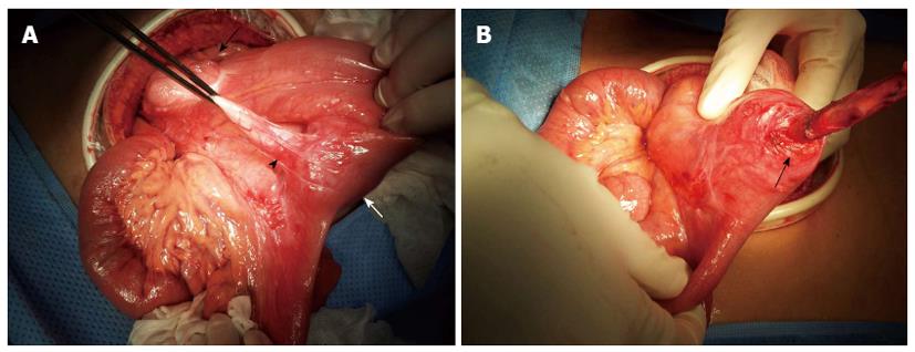
Figure 2 Intraoperative findings.
A: The appendix was attached to the terminal ileum and a firm mass was palpable within the terminal ileum (ileocecal valve, black arrow; appendiceal tip, black arrowhead; palpable mass, white arrow); B: The appendix base was divided and dissection was performed to the lesion to the entry of the appendiceal tip into the terminal ileum (entry site, black arrow).
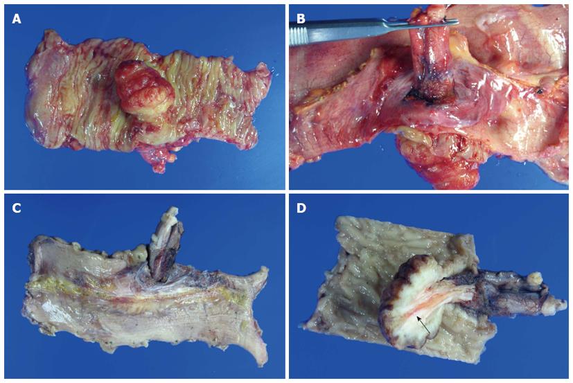
Figure 3 Postoperative gross findings.
A: The polyp size was 3.5 cm × 3.0 cm; B, C: The appendiceal tip was continuous from external to the ileum into the ileal lumen; D: Tissue cross section revealed that the appendix penetrated into the polyp inside the ileum. The appendiceal lumen was identified within the ileum (black arrow).
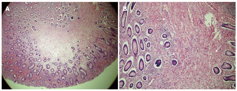
Figure 4 Histopathologic findings (hematoxylin and eosin staining).
A: It was shown hyperplastic epithelium. But there was no dysplasia (× 40); B: Demonstrating the arborizing pattern of smooth-muscle proliferation (white arrow). And these smooth muscle bundles were originated from the muscularis mucosa (× 100).
- Citation: Choi CI, Kim DH, Jeon TY, Kim DH, Shin NR, Park DY. Solitary Peutz-Jeghers-type appendiceal hamartomatous polyp growing into the terminal ileum. World J Gastroenterol 2014; 20(16): 4822-4826
- URL: https://www.wjgnet.com/1007-9327/full/v20/i16/4822.htm
- DOI: https://dx.doi.org/10.3748/wjg.v20.i16.4822


